This 10 year old neutered male Beagle cross was presented for altered behavior. The physical exam was normal. CBC was normal while the blood chemistry revealed moderately elevated SAP and a slightly elevated total protein and slightly elevated globulin. The urinalysis revealed USG of 1.021 and 4+ proteinuria with inactive sediment. ACTH stimulation test was normal.
This 10 year old neutered male Beagle cross was presented for altered behavior. The physical exam was normal. CBC was normal while the blood chemistry revealed moderately elevated SAP and a slightly elevated total protein and slightly elevated globulin. The urinalysis revealed USG of 1.021 and 4+ proteinuria with inactive sediment. ACTH stimulation test was normal.
Case Study
Gastric epithelial tumor and pancreatic cyst in a 10 year old MN Beagle mix dog
Sonographic Differential Diagnosis
Pedunculated gastric mural mass arising from the superficial layers. Differentials include neoplasia (possibly of epithelial origin), granulomatous disease and, less likely, a hematoma. Pancreatic cysts and chronic active pancreatitis likely. Potential abscess. Minimal potential for neoplasia
Image Interpretation
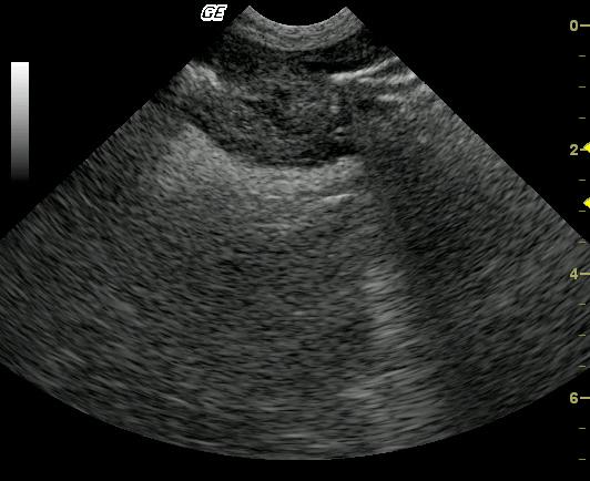
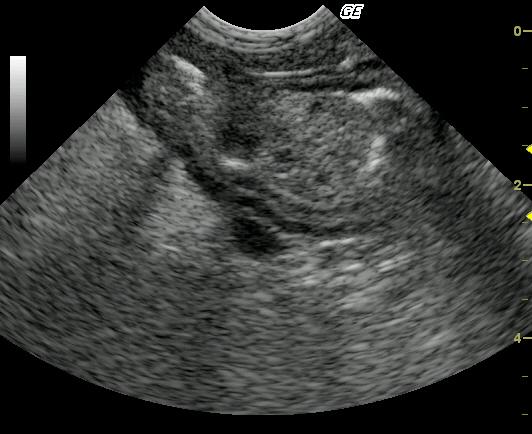
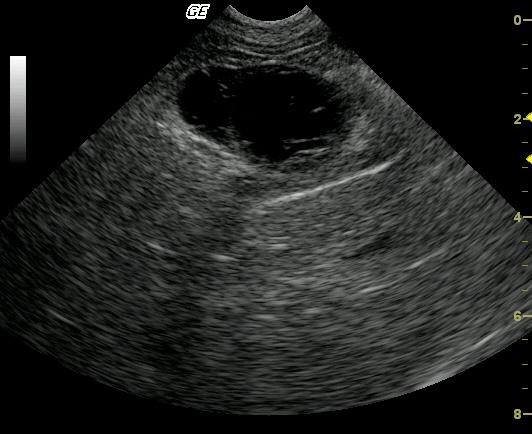
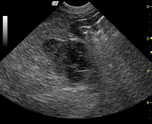
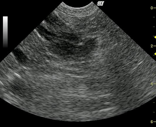
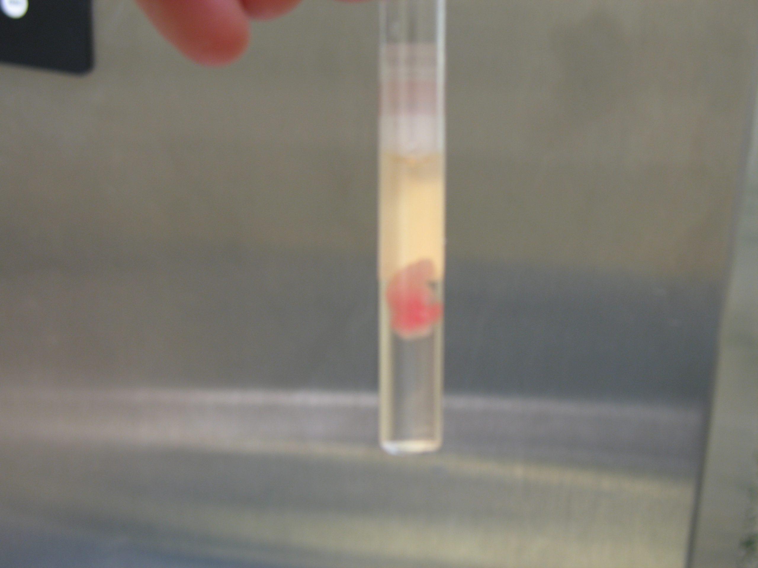
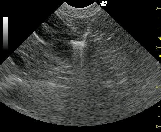
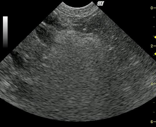
There is asymmetrical thickening of the gastric wall at the level of the pyloric antrum (Image 1). Critical assessment of the wall is difficult due to intervening luminal gas; however, loss of wall layering appears to be present in this image. The mass appears to be protruding into the gastric lumen (image 2). The gas borders the cranial and dorsal margins (left lateral and far field aspects of the mass), supporting a pedunculated type mass predominantly arising from the greater curvature. This is an important finding to recognize, because it suggests complete surgical resection may be possible. In the far field aspect of the image, the muscularis and serosal layers bordering the mass appeared to be uninvolved, suggesting an origin within the more superficial layers. This was confirmed on cytology. Color flow Doppler indicates tissue protrusion into the lumen as opposed to attached foreign material or food that may appear similarly (Video 1). US-guided FNA of the gastric mass can be seen in video 2. A mixed hypoechoic cystic lesion at the base of the pancreatic body is present(Image 3). Hypoechoic tissue with hyperechoic remodeled and potentially inflamed fat is noted, US-guided drainage was performed (Video 3 show post drainage) with enrofloxacin injected due to the flocculent macroscopic nature of the fluid to combat any sequestered infection that may be present.
DX
Well-differentiated gastric epithelial tumor, pancreatic cyst.
Outcome
The owners decided to not pursue further therapeutic measures. The patient was treated medically.
Comments
The pancreatic lesion was largely incidental. There was a concurrent well-differentiated gastric carcinoma. The decision to infuse the lesion with enrofloxacin was made at the time of the sonogram due to the slight inflammatory aspect of the fluid (see image 6) and presence of the granulation bed around the lesion. This procedure was a palliative intervention on the part of the sonographer (EL) that is not supported in the literature.
Clinical Differential Diagnosis
SAP elevations – Vacuolar hepatopathy, cholangiohepatitis, neoplasia. Isosthenuria – glomerulonephritis, chronic renal disease. Proteinuria – protein losing nephropathy (familial nephropathy associated with structural abnormalities of the glomerular capillary wall, glomerular amyloidosis, and immune complex glomerulonephritis).
Sampling
22 and 20-gauge US-guided FNA revealed well differentiated gastric epithelial tumor with minimal atypia (Video 2). The corkscrew technique was employed during sampling after the simple jab technique failed to provide adequate samples when sprayed on the slide. US-guided drainage of the pancreatic cyst revealed a low grade inflammatory modified transudate.
UA Specific Gravity Range
1.021
Video
Patient Information
Patient Name :
Sammy Y
Gender :
Male, Neutered
Species :
Canine
Type of Imaging : Ultrasound
Book :
yes
Status :
Complete
Liz Wuz Here :
Yes
Code :
04_00101
Clinical Signs
- "Not Doing Right"
Images
Blood Chemistry
- Alkaline Phosphatase (SAP), High
- Globulin, High
- Total Protein, High
Clinical Signs
- "Not Doing Right"
Urinalysi
- Protein Present
- Sediment, inactive