Esophageal stenosis, segmental megaesophagus and esophageal dysmotility in a 2 year old FS Corgi dog

Case Study

Esophageal stenosis, segmental megaesophagus and esophageal dysmotility in a 2 year old FS Corgi dog

History of chronic vomiting, regurgitation, aspiration pneumonia and suspected megaesophagus or swallowing deficits. Physical exam: very underweight

History of chronic vomiting, regurgitation, aspiration pneumonia and suspected megaesophagus or swallowing deficits. Physical exam: very underweight

Image Interpretation

Barium Swallow Fluoroscopy: The cervical, cranial thoracic and mid thoracic esophagus presented severe segmental dilatation. Cranial to the diaphragm constrictive narrowing of the caudal thoracic esophagus was noted. Vigorous secondary peristaltic waves were not able to empty the dilated esophagus beyond the stenosis as the narrowing persisted throughout all phases of swallowing/peristalsis. Only scant amounts of the barium soaked meal were able to pass the stenosis and enter the stomach. The secondary peristaltic waves appeared to descend the length of the esophagus incompletely.

DX

The fluoroscopic findings are compatible with a caudal thoracic esophageal stenosis and secondary segmental megaesophagus. The findings further suggest esophageal dysmotility.

Outcome

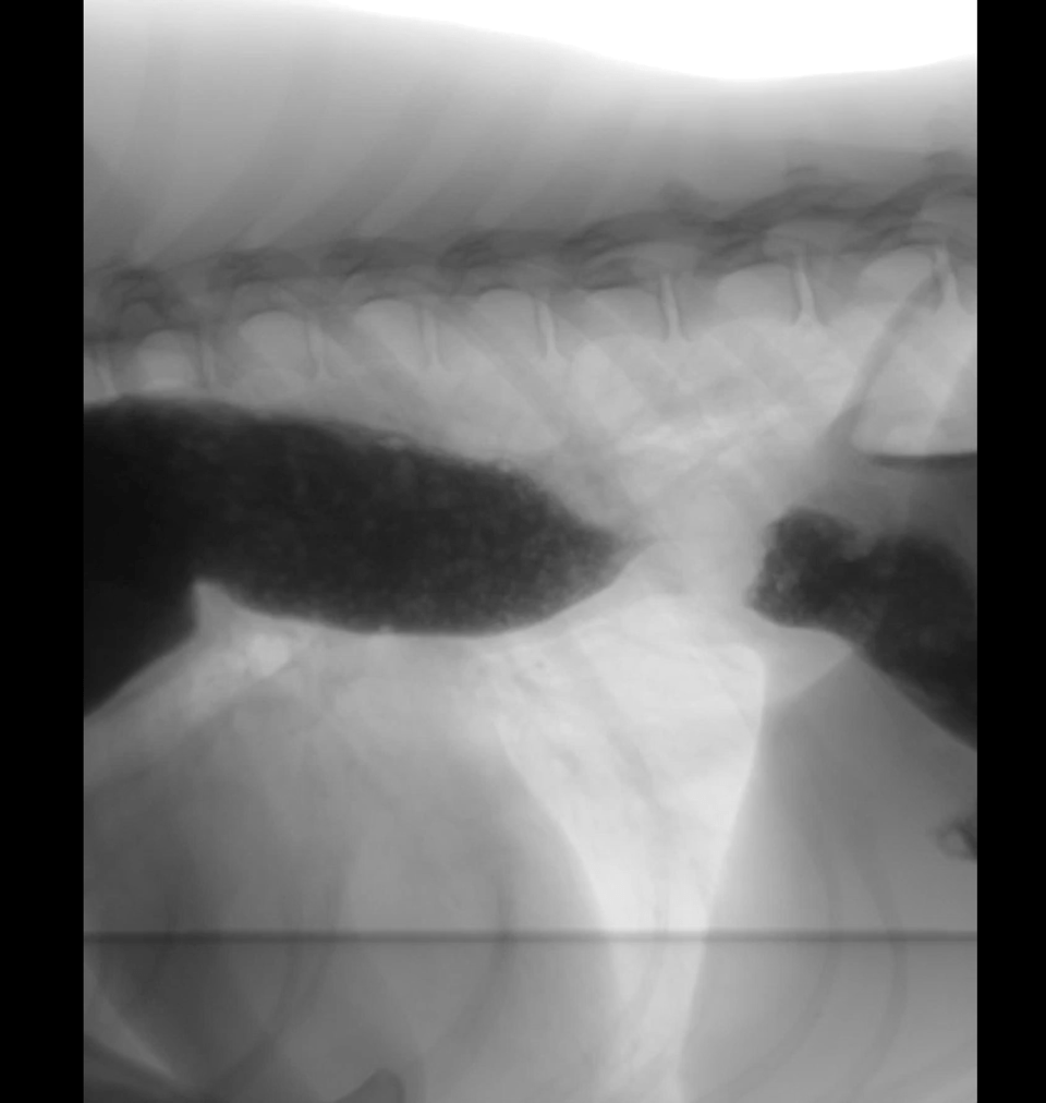
The cause of the stenosis may be mechanical, such as a stricture, or functional, such as lower esophageal sphincter achalasia or asynchrony, either.
Potential causes of stricture include traumatic, such as foreign bodies or chemical irritation, and congenital. Esophageal dysmotility disorders overall are lower for potential as the position of the obstruction cranial to the diaphragm does not match the regular anatomy.
The generalized esophageal dysmotility likely is a late sequela to the chronic dilation and may be irreversible. The prognosis is guarded.
Endoscopy was recommended to plan therapeutic approach such as interventional balloon dilation or surgery.

Clinical Differential Diagnosis

megaesophagus or swallowing deficits

Patient Information

Patient Name : Pistol, Neel Veterinary Hospital
Gender : Female, Spayed
Species : Canine
Type of Imaging : Ultrasound
Status : Complete

Clinical Signs

  • Vomiting

Exam Finding

  • Weight loss

Images

bildschirmfoto_2015-09-02_um_17

Clinical Signs

  • Vomiting
Skip to content