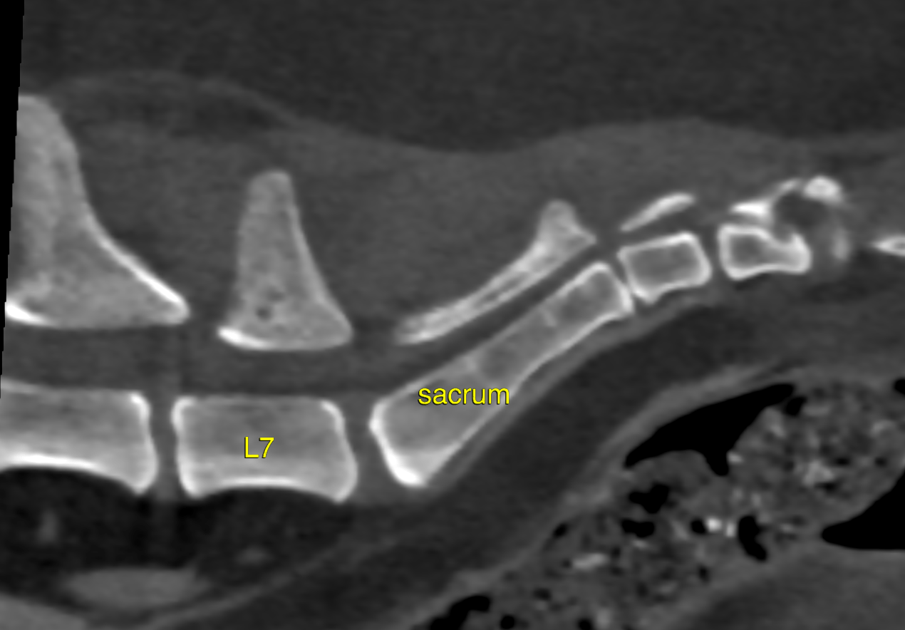
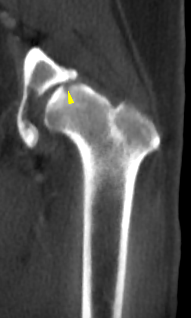
This 9 year old FS canine mix presented with a history of difficulty walking and painful in her hind end. Rads were taken and an osteoarthritis was noted in her left hip. She began taking Tramadol and Rimadyl. She was seen for a follow-up pne month later and radsshowed changes in the left femoral head. CT was done in order to help rule out trauma, femoral head and neck necrosis, or neoplasia.
Physical Exam: Acute lameness (left side)