CT – Mild intervertebral disc protrusion and congenital scoliotic malformation in a 5 year old MN English Bulldog

Case Study

CT – Mild intervertebral disc protrusion and congenital scoliotic malformation in a 5 year old MN English Bulldog

This 5 year old MN English Bulldog presented with severe neck pain beginning 12/18. Pulled aggressively on leash 12/17, but symptoms did not present for 24 hours.

CBC/Chem/U/A not done


This 5 year old MN English Bulldog presented with severe neck pain beginning 12/18. Pulled aggressively on leash 12/17, but symptoms did not present for 24 hours.

CBC/Chem/U/A not done


Image Interpretation

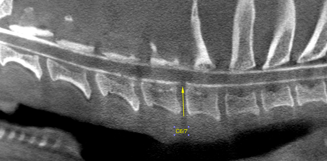
CT of the spine, plain and myelogram- The computed tomography show mild herniation of the disc C6/7 which is located centrally within the ventral epidural space. The ventral contrast column is obliterated. The lateral and dorsal columns as well as the cross sectional area of the spinal cord are maintained. There is no spinal cord displacmement or compression.

Mild scoliosis of the mid thoracic spine is noted from T4 to T8. This is not paralleled by compressive myelopathy.

The distribution of the contrast media within the epidural space is non uniform level with the thoracolumbar and lumbar spine which is likely to be a function of the low volume of contrast here.

There is mild bilateral hip dysplasia with mild to moderate coxofemoral osteoarthritis. Clinical relevance is questionable at this point. The esophagus reveals mild generalized dilation with fluid accumulation under general anesthesia. Consider symptomaitc treatment of gastritis in case of clinical signs of reflux esophagitis/gastritis.

DX

mild intervertebral disc protrusion C6/7 with no relevant compressive myelopathy at this point. Moreover there is a congenital scoliotic malformation of the mid thoracic spine which is an incidental finding here.

Outcome

A compressive myelopathy or traumatic injury requiring surgical intervention is not encountered. Symptomatic conservative treatment and monitoring of the neurological status is recommended.

Note that the severe neck pain experienced by the patient may be a function of the aforementioned disc protrusion. But other pathologies such as traumatic disc extrusions (high impact/low volume extrusion), which frequently occur after vigourous exercise or handling as indicated in the history here, or spinal cord infarction are possible differential diagnoses as they are not necessarily apparrent on a myelogram.

Patient Information

Patient Name : Oscar Hiers
Gender : Male, Neutered
Species : Canine
Type of Imaging : Ultrasound
Status : Complete
Liz Wuz Here : Yes

Clinical Signs

  • Pain

Images

bildschirmfoto_2015-12-23_um_11

Clinical Signs

  • Pain
Skip to content