This Standard Schnauzer dog presented with chronic hip pain. Rads showed lysis on left hip. Intermittent lameness left hind.
This Standard Schnauzer dog presented with chronic hip pain. Rads showed lysis on left hip. Intermittent lameness left hind.
This Standard Schnauzer dog presented with chronic hip pain. Rads showed lysis on left hip. Intermittent lameness left hind.
This Standard Schnauzer dog presented with chronic hip pain. Rads showed lysis on left hip. Intermittent lameness left hind.
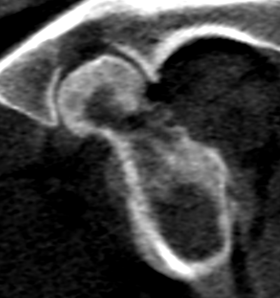
CT of the hips – There are multifocal geographic osteolytic zones within the left femoral head and neck.
The trabecular pattern of the bone is irregular. The subchondral bone plate presents
multifocal sclerotic zones. There are solid periosteal reactions circumferential to the
left femoral metaphysis with mildly unsharp surface. Marked medullary sclerosis is
noted extending from the metaphysis into the proximal third of the diaphysis of the left
femur.
A subtle remnant of the former femoral head physis is recognized.
Mild irregularity of the left hip joint space is noted. Emerging osteophytes are seen at
the left acetabular rim.
There is no soft tissue swelling nor increased contrast enhancement within the
surrounding soft tissues.
There is generalized osteopenia with marked cortical thinning of the left femur.
The volume of the left thigh musculature is moderately reduced.
The left medial iliac lymph node reveals mild symmetric enlargement with maintained
short-to-long-axis ratio and contrast enhancement pattern.
The right femur and hip joint are within normal limits.
The findings are compatible with avascular necrosis of the left femoral head & neck
within the late avascular phase. The femoral head is prone to collapse.
The differential diagnoses bone neoplasia and juvenile hematogenous osteomyelitis are
very unlikely.
Femoral head and neck ostectomy plus pathohistological examination is recommended
followed by physical therapy.