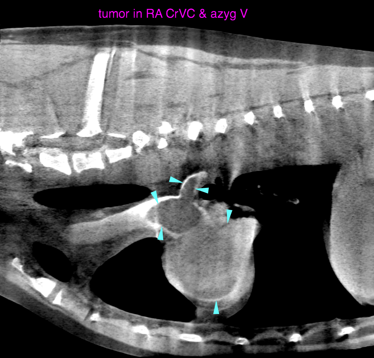
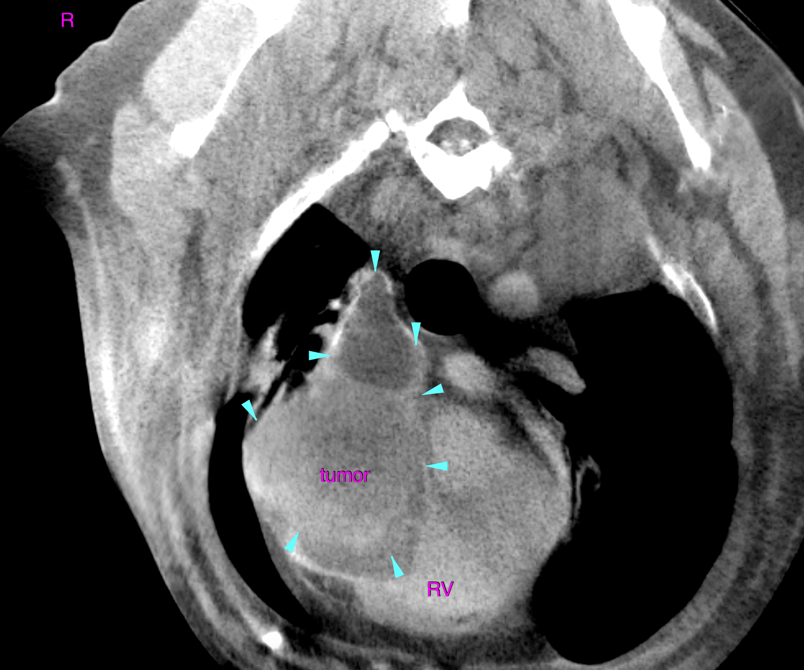
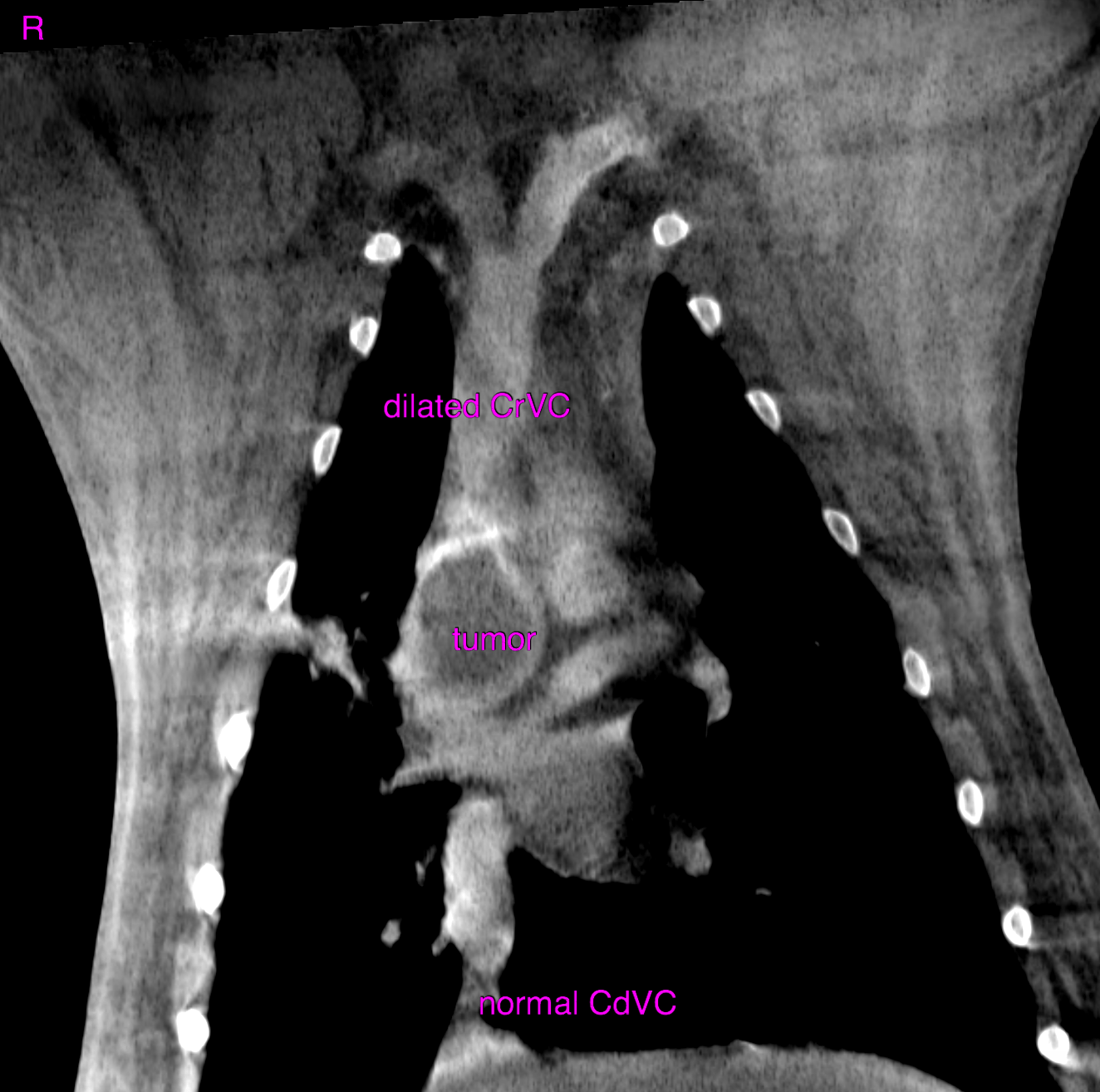
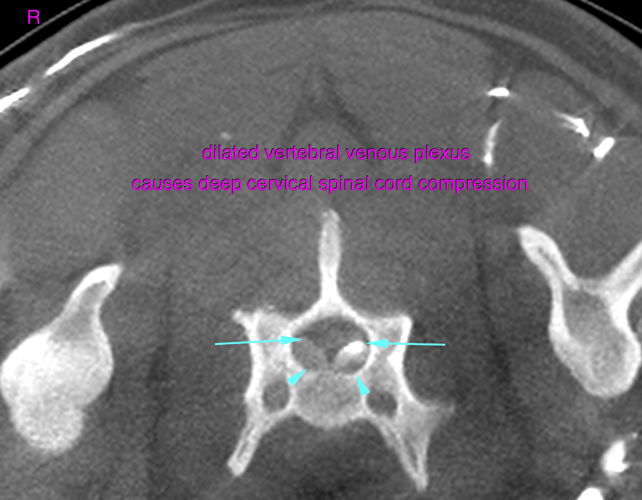
This 8 year old M mixed breed dog presented with pleural effusion, facial swelling . Fluid analysis showed modified transudate. On echo a 4.5 cm suspected soft tissue mass is identified at the heart base.
This 8 year old M mixed breed dog presented with pleural effusion, facial swelling . Fluid analysis showed modified transudate. On echo a 4.5 cm suspected soft tissue mass is identified at the heart base.