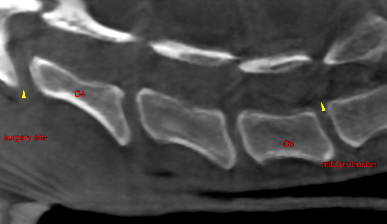
This 11 year old MN Border Collie dog presented with lameness of the right front and “wobbliness” of 24 hour duration. CBC/Chem wnl.
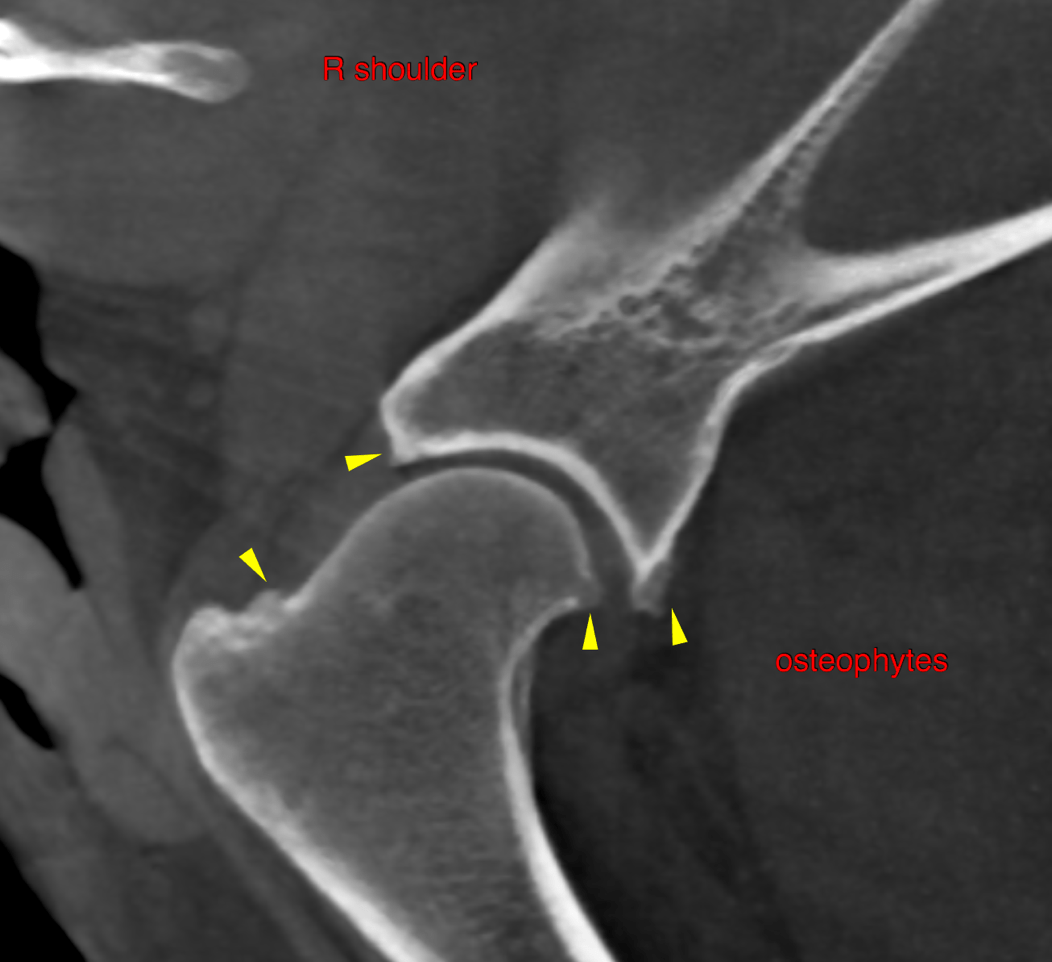
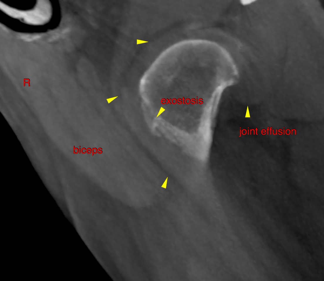
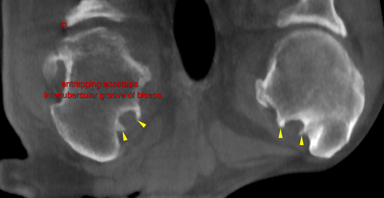
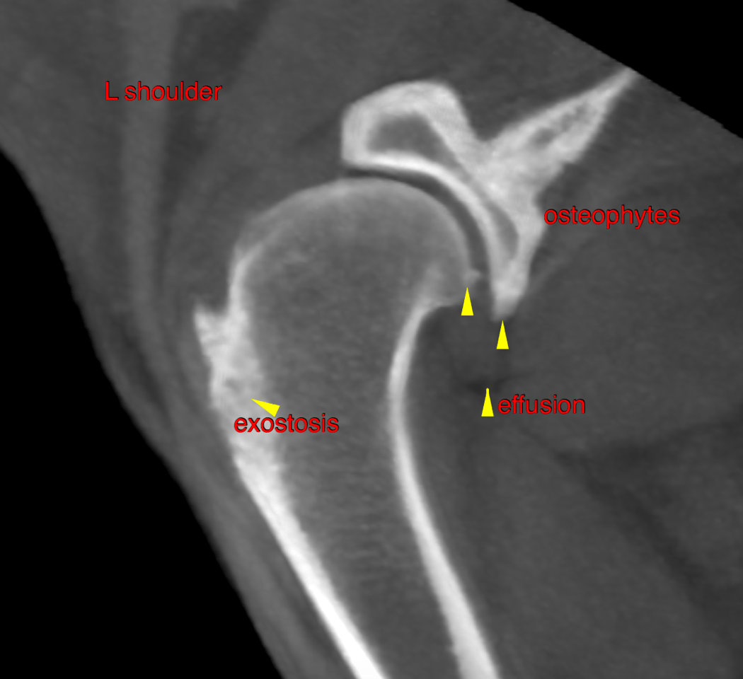
Physical exam – cervical myelopathy (C3-4 disc surgery today) and pain on right shoulder flexion. Post-op CT to determine cause of right shoulder pain.
This 11 year old MN Border Collie dog presented with lameness of the right front and “wobbliness” of 24 hour duration. CBC/Chem wnl.
Physical exam – cervical myelopathy (C3-4 disc surgery today) and pain on right shoulder flexion. Post-op CT to determine cause of right shoulder pain.