CT – Bilateral Parathyroid Hyperplasia consistent with Hyperparathyroidism

Case Study

CT – Bilateral Parathyroid Hyperplasia consistent with Hyperparathyroidism

The patient has a history of persistent hypercalcemia.

The patient has a history of persistent hypercalcemia.

Image Interpretation

CT of the thorax and abdomen, plain and post contrast series – There were bilateral signs of moderate hip dysplasia with secondary osteoarthrosis.
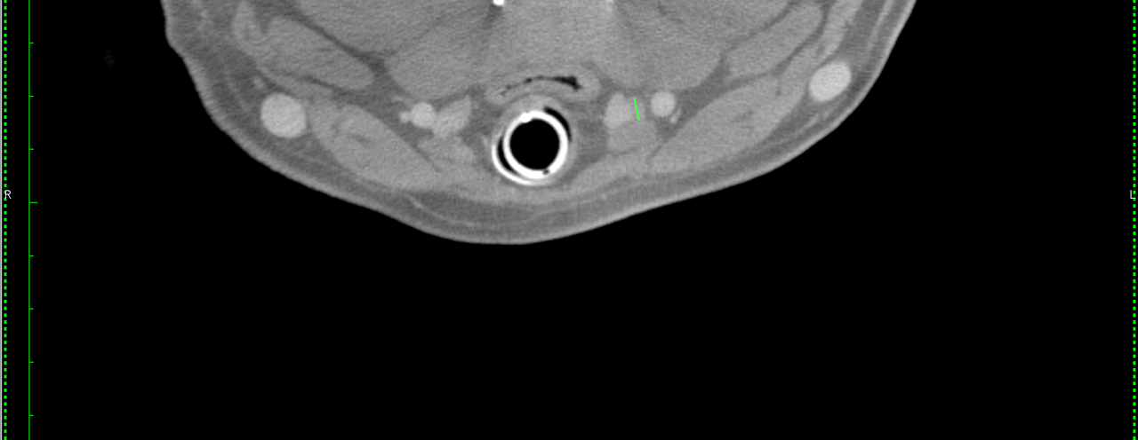
There was mild bilateral generalized enlargement of the external parathyroid glands up to a maximum diameter of 4mm.
The thyroid glands were regular in shape, size, attenuation and enhancement behavior.
The internal parathyroid glands were not seen.
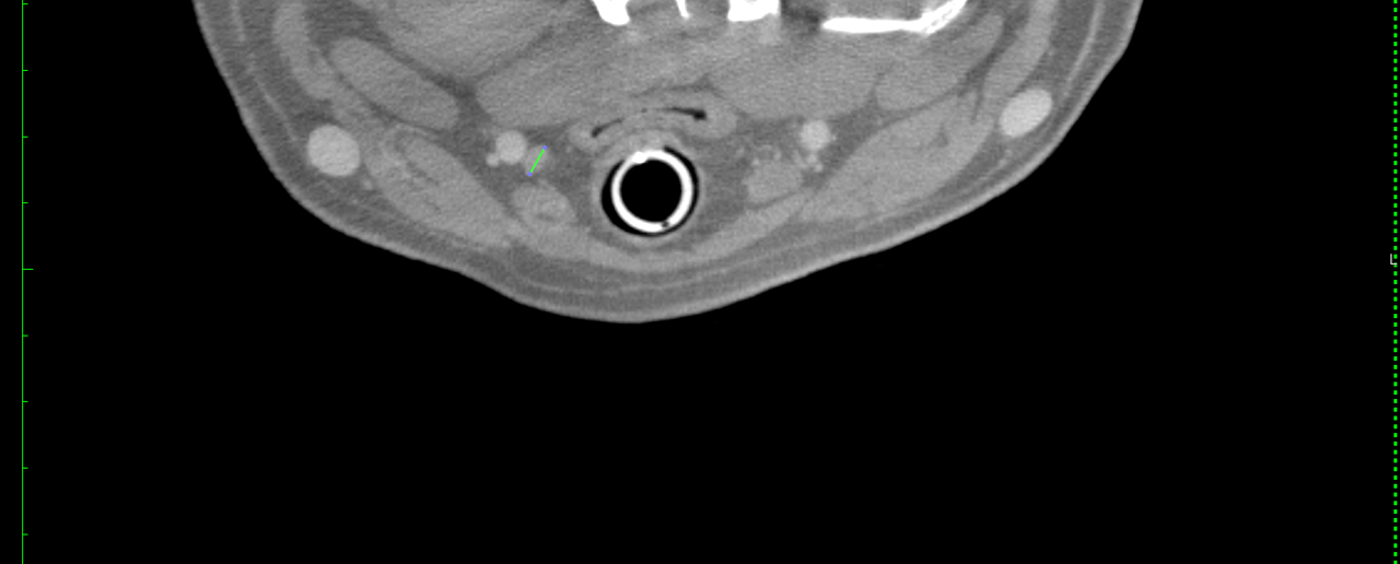
The mediastinal and abdominal lymph nodes were within normal limits.
The medial iliac and hypogastric lymph nodes were within normal limits.
The anal sacs were not included in the scan.
There were moderate signs of pressure related atelectasis in the dependent portion of the lungs.
The spleen was large and presented rounding of the splenic head which likely was a function of anesthesia related congestion.

DX

The findings suggest mild bilaterally symmetric parathyroid hyperplasia which may be consistent with secondary hyperparathyroidism (renal versus nutritional). Primary hyperparathyroidiIncidental hip dysplasia and coxarthrosis was noted bilaterally.sm is a potential too. Incidental hip dysplasia and coxarthrosis was noted bilaterally.

Outcome

Calcium, Phosphorus and Parathormone levels should be checked. Rule out chronic renal failure. Please note the degree of enlargement is minor and equivocal here.
The findings associated with the spleen likely are related to anesthesia. A diffuse neoplastic round cell infiltrate – such as lymphoma – cannot be ruled out for the spleen and liver. Consider fine needle aspiration of the liver and spleen since lymphoma is one potential underlying cause of malignant hypercalcemia. Rule out anal gland adenoadenocarcinoma which may be associated with parathormone like substance production.

Patient Information

Patient Name : Duke Wollney
Type of Imaging : Ultrasound
Status : Complete

Images

bildschirmfoto_2015-08-05_um_20bildschirmfoto_2015-08-05_um_20
Skip to content