A 9-year-old NM Gordon Setter was presented for evaluation of ocular disease characterized by conjunctivitis and increased intraocular pressure. Weight loss was present on physical examination. CBC and serum biochemistry were within reference range.
A 9-year-old NM Gordon Setter was presented for evaluation of ocular disease characterized by conjunctivitis and increased intraocular pressure. Weight loss was present on physical examination. CBC and serum biochemistry were within reference range.
Right eye – chorioretinitis with iriditis and posterior lens luxation. Left eye – the iris appears slightly thickening, however this may be normal for this patient. However, preventive treatment is recommended regardless. Emergency therapy for glaucoma is recommended. Approximately 70% of the normal eye in case of glaucoma will develop glaucoma within 9 months; therefore preventive therapy to the left eye may be appropriate. It is debatable whether the right eye is salvageable in this case.
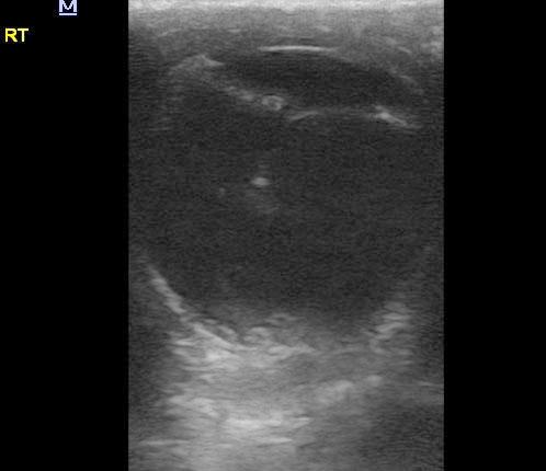
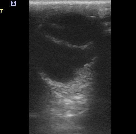
Left eye revealed normal retina. Anechoic fluid in the posterior and anterior chambers. Lens was in proper position. Periocular tissues appeared uniform. No evident pathology. The right eye revealed minor posterior chamber precipitate. Retina appears thickened and irregular as does the iris in the right eye. Posterior luxation of the lens is present.
None
Glaucoma, neoplasia, lens-luxation, chorio-retinitis, uveitis
None