A 5-year-old NM Bernese Mountain Dog was presented for evaluation of coughing.
Sonographic Differential Diagnosis
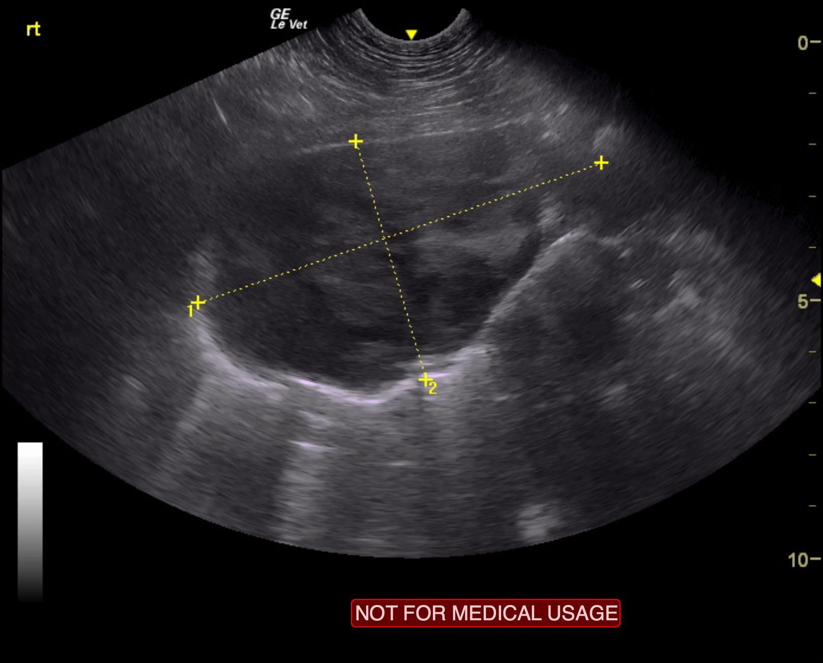
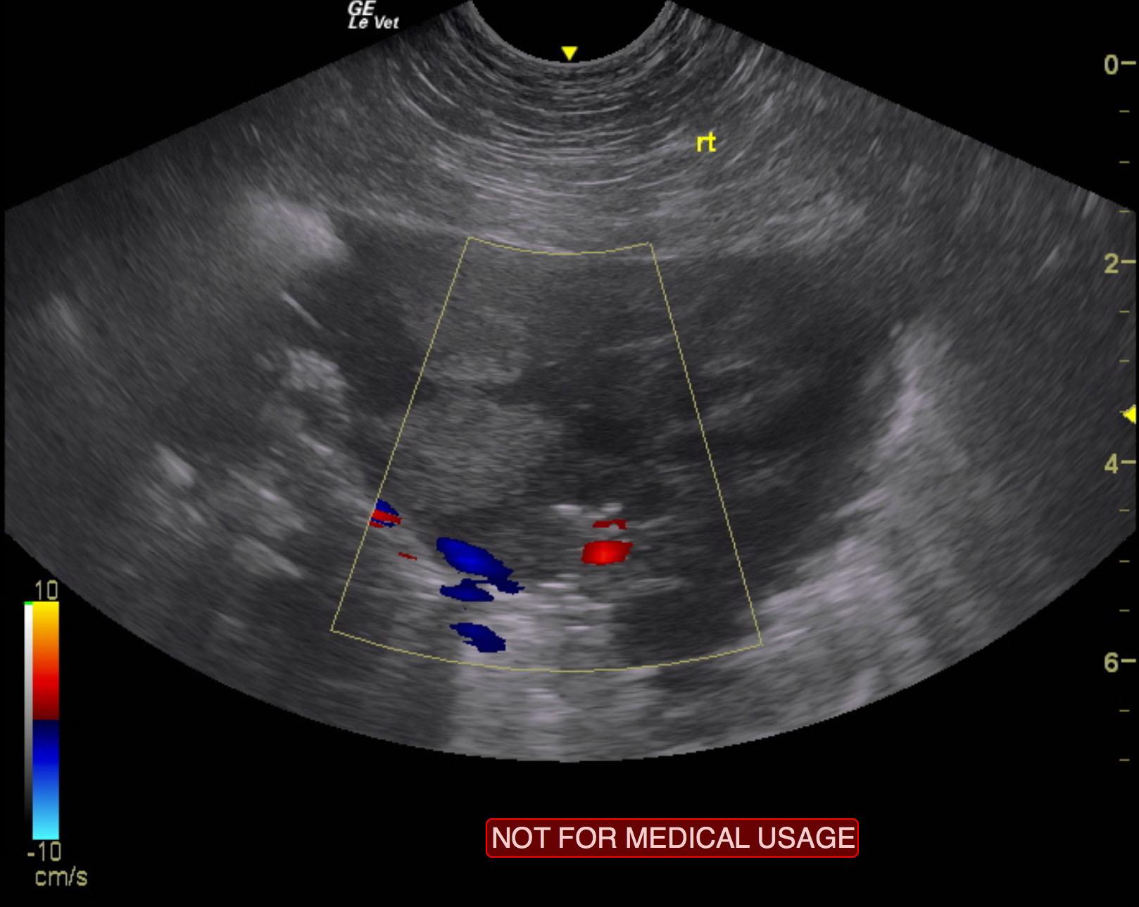
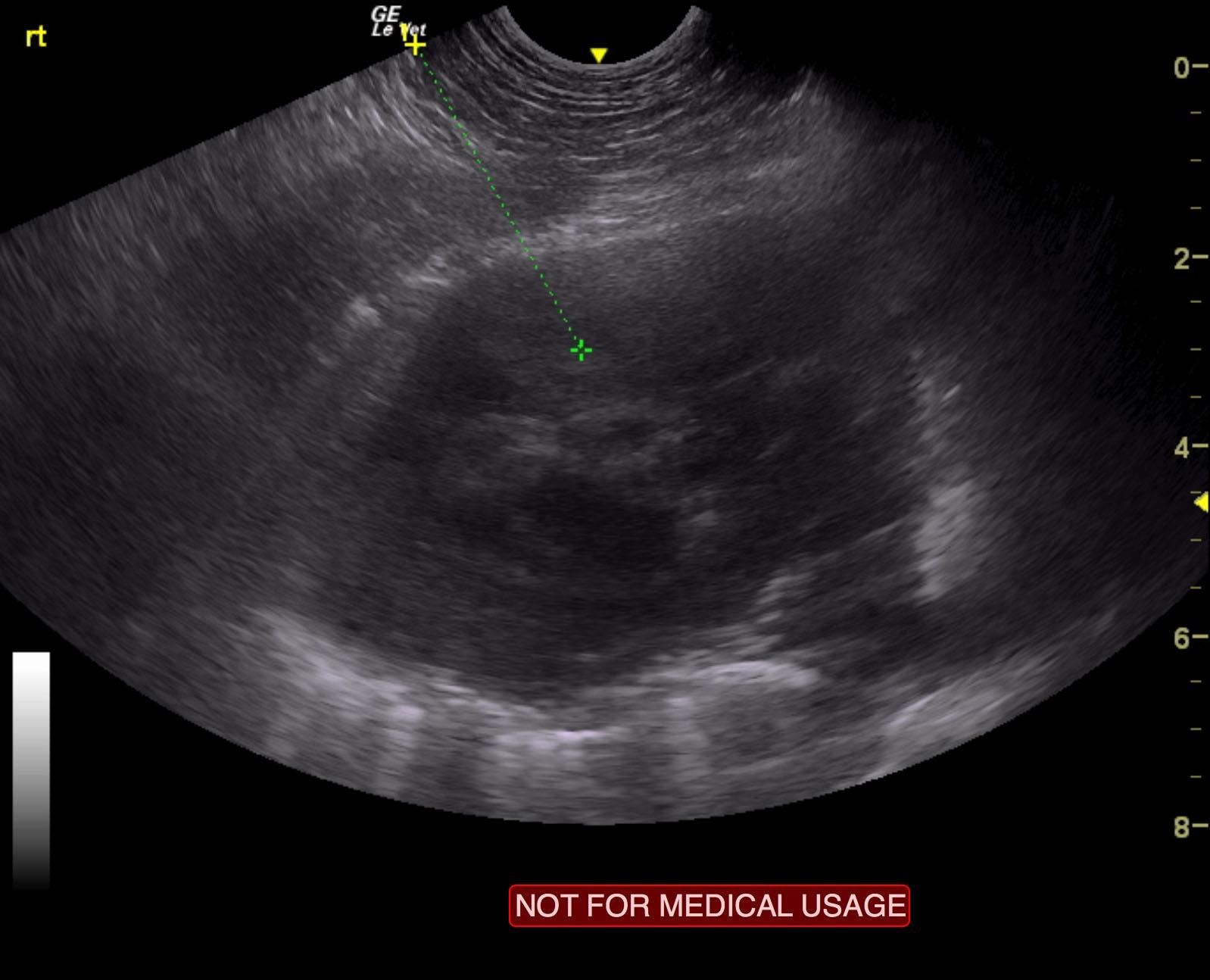
Primary lung mass. Strong suspicion for sarcoma or other round cell neoplasia.
Passive obstructive pattern of the vena cava causing minor passive congestion pattern of the liver. FNAs were performed of the lung mass without complication.
Image Interpretation
Right lung presented a cavitated, mixed, hypoechoic mass that was strongly suggestive for sarcoma with peripheral air entrapment indicating lung origin. It appears to be lobar in nature. However, the extent of the mass was ill defined throughout the lung. Passive obstructive pattern of the vena cava causing minor passive congestion pattern of the liver