09_00174 Gus S Prostatic cyst

Case Study

09_00174 Gus S Prostatic cyst

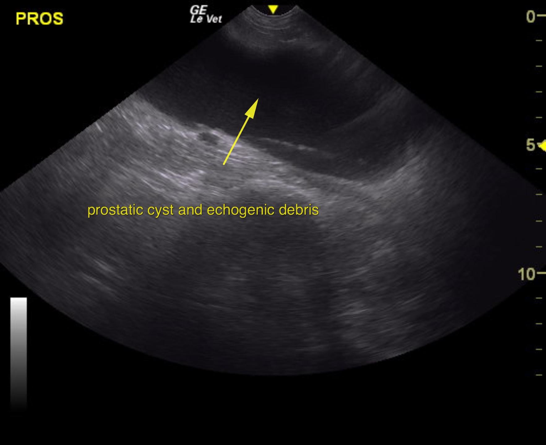
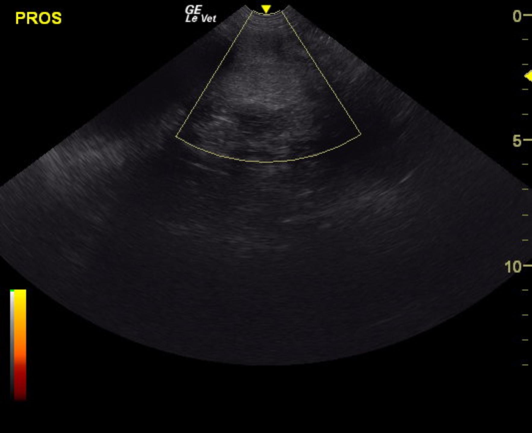
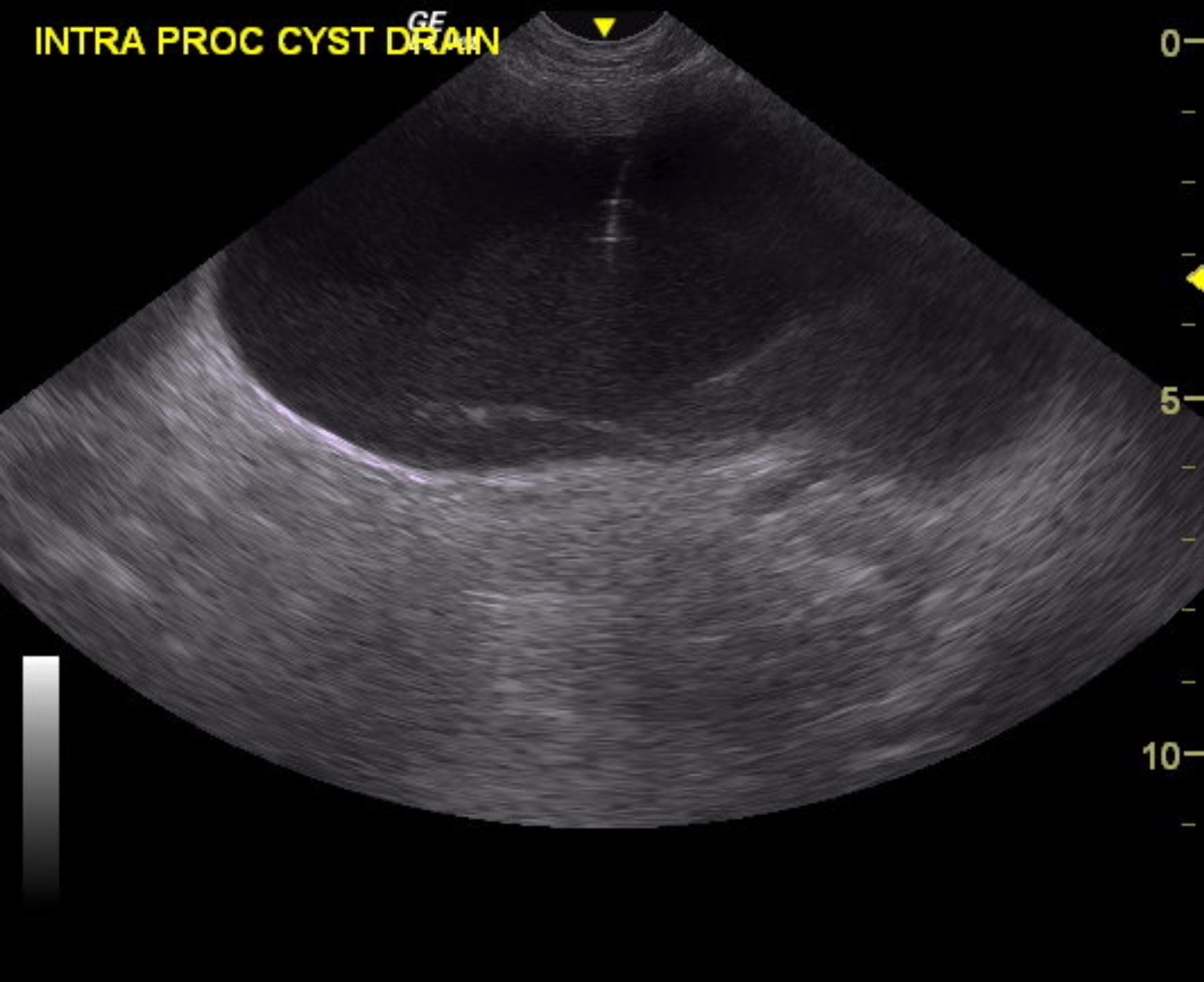
A 7-year-old intact male Boxer was presented for evaluation of tenesmus and hematochezia with normal urinalysis, CBC, and serum biochemistry. Prostatomegaly was present on rectal palpation. Survey radiographs showed a mass caudal to the urinary bladder.


Sonographic Differential Diagnosis

Prostatic cyst. Ultrasound-guided drainage and neuter in this patient is recommended.

Image Interpretation

A large prostatic cyst was noted and occupied the pelvic inlet. The remaining parenchymal portion of the prostate measured approximately 4.0 cm and was uniform, most consistent with BPH. The testicles were imaged and found to be uniform with no evident pathology.

DX

Prostatic cyst

Outcome

Post drainage and after antibiotic injection, the prostatic cyst measured approximately 3 x 2 cm.

Clinical Differential Diagnosis

Prostate – benign hyperplasia, prostatitis, abscessation, para-prostatic cyst, neoplasia

Sampling

Cyst was drained. No information regarding fluid appearance or culture was provided.

Patient Information

Gender : Male, Intact
Species : Canine
Type of Imaging : Ultrasound
Status : Complete

Clinical Signs

  • Fresh Blood in Stool
  • Tenesmus

Images

as2gus3gus2gus4

Clinical Signs

  • Fresh Blood in Stool
  • Tenesmus
Skip to content