09_00173 Zachary M Prostatitis, possible abscess

Case Study

09_00173 Zachary M Prostatitis, possible abscess

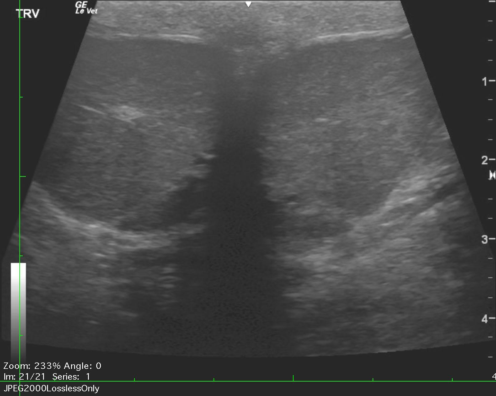
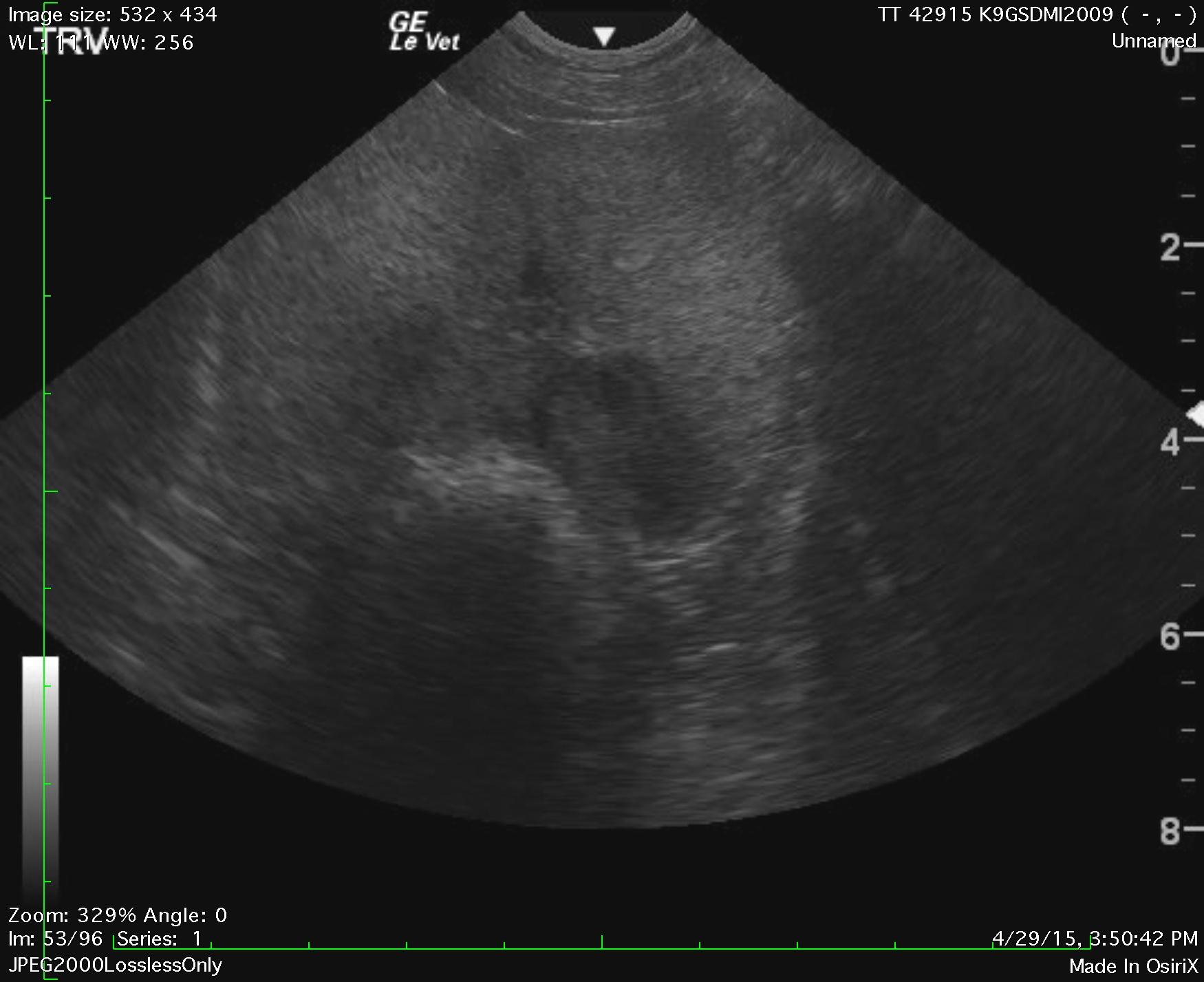
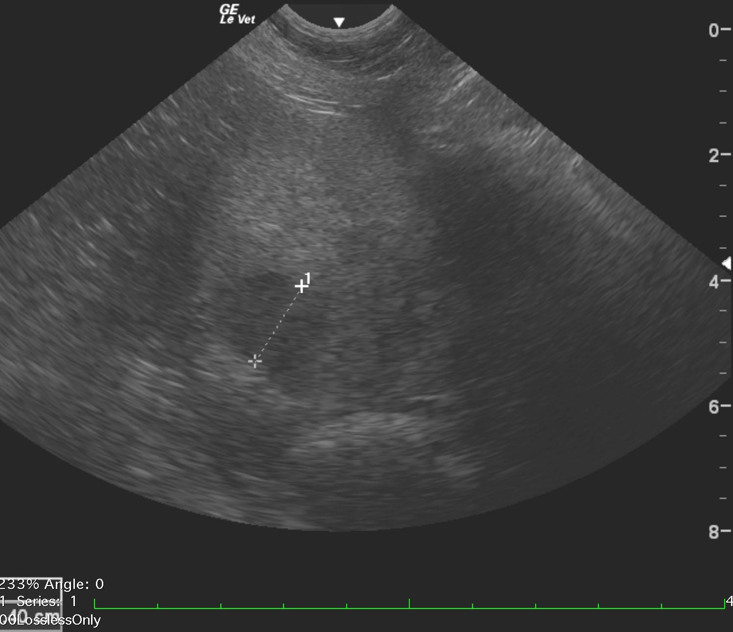
A 6-year-old intact male German shepherd was presented for evaluation of intermittent drops of blood at the prepuce. Physical examination was unremarkable. Free flow urinalysis showed leukocytes and possible cocci but culture and sensitivity yielded no growth.


Sonographic Differential Diagnosis

Prostatitis. Potential abscessation.

Image Interpretation

The testicles were imaged and found to be uniform. There was no evidence of pathology. The prostate in this patient was enlarged with a 1.4 cm nodule at the lateral aspect of the right lobe of the prostate. The prostatic nodule may represent an abscess given the echogenicity. Focal capsular expansion was also noted. The prostate itself presented parenchymal striations and micropunctate changes. This is consistent with prostatitis.

DX

Prostatitis, possible abscess

Outcome

None

Clinical Differential Diagnosis

Prostate – benign prostatic hyperplasia, prostatitis, abscessation, neoplasia
Bladder – urolith, neoplasia
Urethra – lith, neoplasia

Sampling

None

Patient Information

Gender : Male, Intact
Species : Canine
Type of Imaging : Ultrasound
Status : Complete

Exam Finding

  • Bleeding from penis

Images

zachery_merts_prostatitis_abscess_testicleszachery_merts_prostatitis_abscess_1zachery_merts_prostatitis_abscess

Urinalysi

  • Bacteria Present
  • Culture negative
  • WBCs Present
Skip to content