06_00335 Marmalade B Bladder mass

Case Study

06_00335 Marmalade B Bladder mass

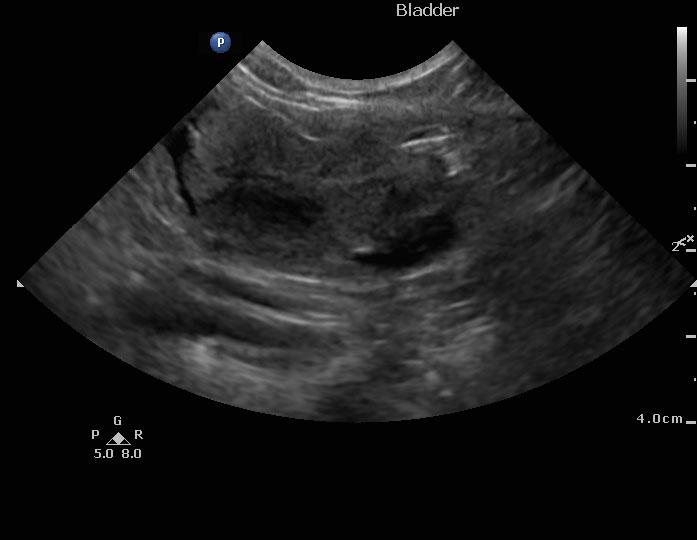
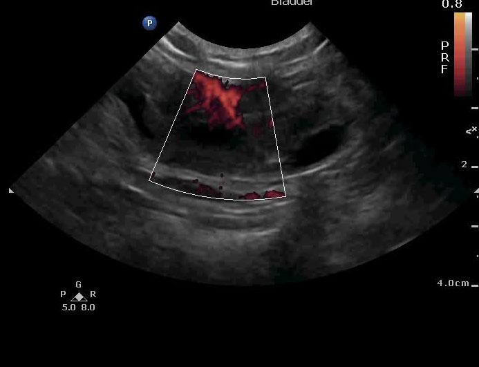
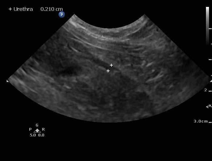
A 9-year-old SF DSH was presented for evaluation of hematuria. A bladder urolith had been diagnosed on survey radiographs that had been dissolved with U/R diet. The only abnormality on urinalysis was the presence of Staphylococcus pseudomonas. CBC and serum biochemistry were within reference range.


Sonographic Differential Diagnosis

Bladder mass. Urethral polyps.
Debulking effort may prove fruitful in long-term management in this case. However, clean resection is unlikely. There was no overt evidence of metastatic disease.

Image Interpretation

A 2.0 x 1.5 cm bladder mass was noted in this patient. The mass was highly vascular and was deriving from the ventral wall with kissing lesions on the dorsal wall. However, early seeding into the urethra may be an issue given the minor, echogenic polypoid changes noted. The pelvic urethra was also dilated and measured 0.25 cm.

DX

Bladder mass

Outcome

None

Clinical Differential Diagnosis

Bladder – interstitial cystitis, neoplasia, chronic cystitis, uroliths, polyploid cystitis, granulomatous cystitis
Kidney – neoplasia, renolith, pyelonephtitis

Sampling

None

Patient Information

Gender : Female, Spayed
Species : Feline
Type of Imaging : Ultrasound
Status : Complete

Clinical Signs

  • Hematuria

Images

120215_mb_allcreatures_bell_20151202_145309_0001vlcsnap-2016-01-31-10h28m54s182120215_mb_allcreatures_bell_20151202_145309_0005

Clinical Signs

  • Hematuria

Urinalysi

  • Bacteria Present
Skip to content