A 7-year-old SF DMH that was presented for dentistry was diagnosed with hydronephrosis. Physical examination, CBC, and serum biochemistry showed no significant abnormalities. Urinalysis showed normal SG, granular cast, and occasional renal cells.
A 7-year-old SF DMH that was presented for dentistry was diagnosed with hydronephrosis. Physical examination, CBC, and serum biochemistry showed no significant abnormalities. Urinalysis showed normal SG, granular cast, and occasional renal cells.
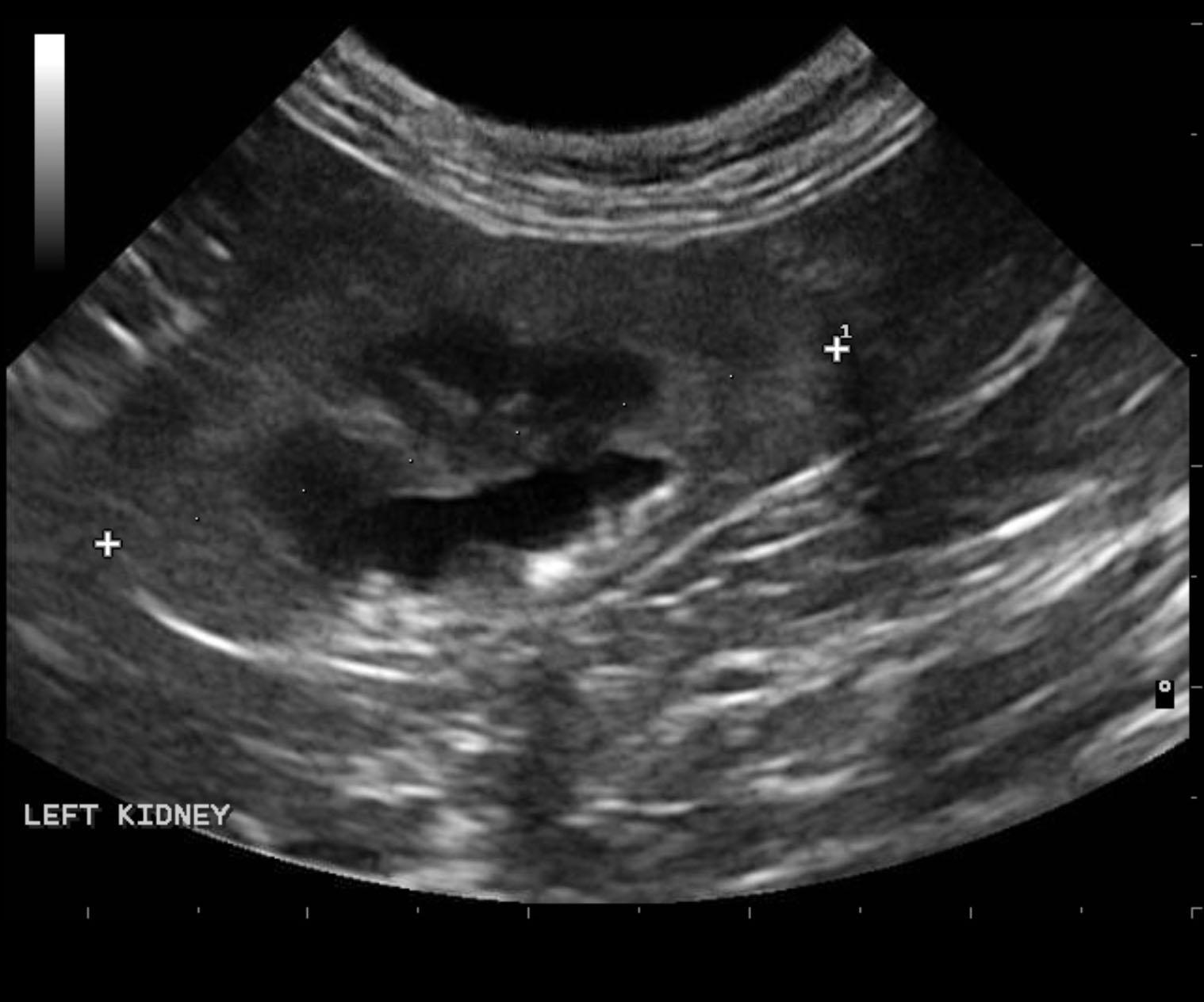
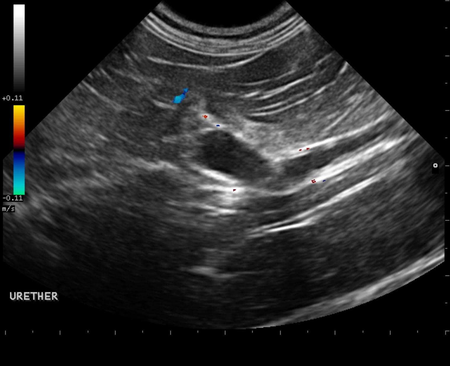
Left ureteral lithiasis with full obstruction. Moderate hydronephrosis.
Multiple left ureteral calculi and left renal pelvic calculi.
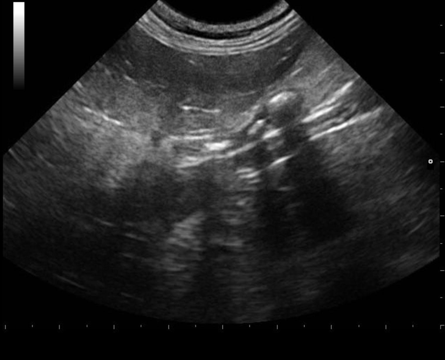
Minor degenerative changes in the right kidney.
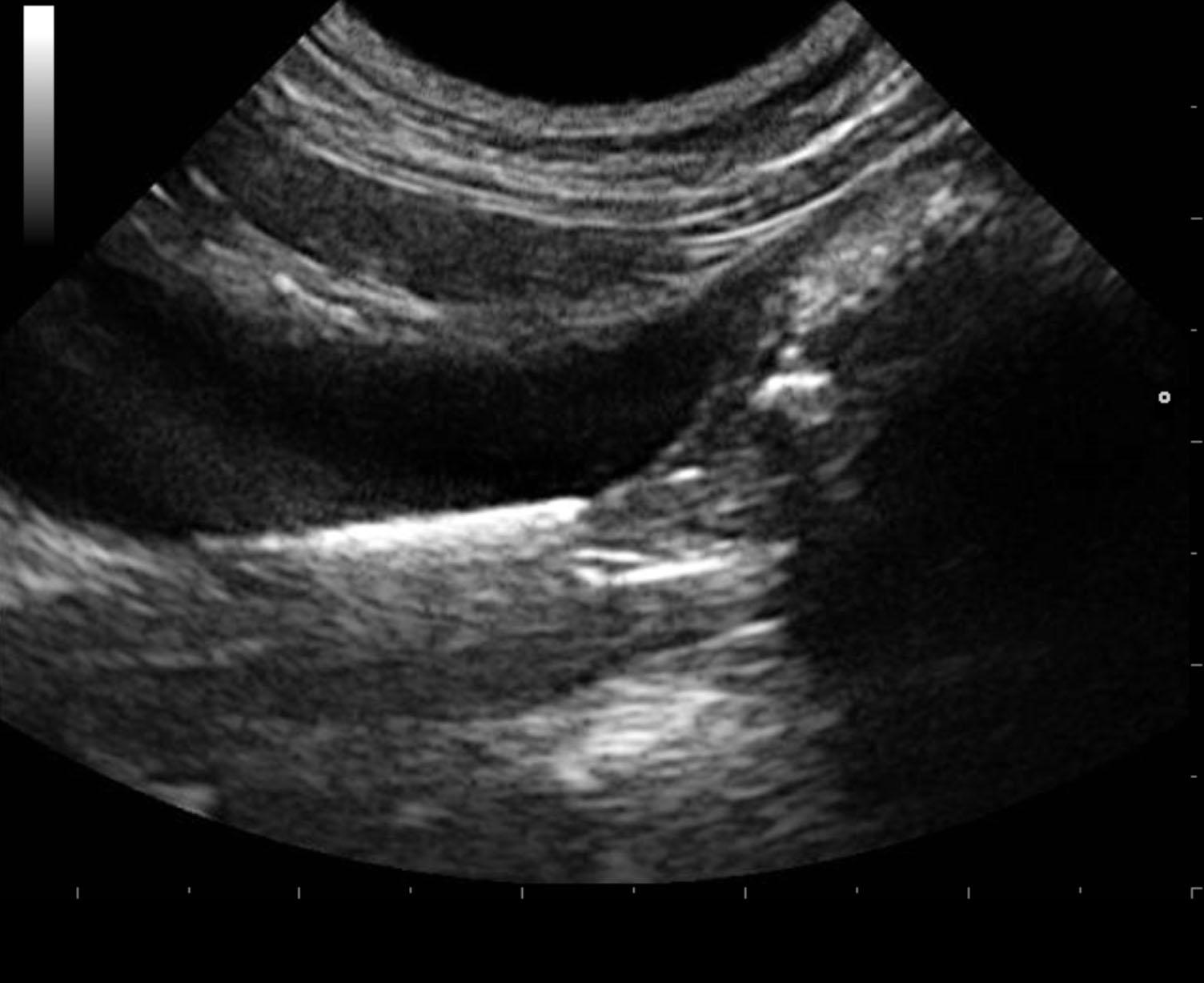
The urinary bladder presented a small amount of sand accumulation measuring approximately 1.0 cm. The bladder wall itself was unremarkable. The left kidney presented moderate hydronephrosis measuring approximately 2.0 cm and 3.4 cm in length with pelvic calculus measuring 0.3 cm. The proximal ureter was dilated at 0.98 cm with ureteral calculus noted at 0.46 cm. This was likely too large to pass into the bladder. The proximal ureter was dilated and mildly thickened. Multiple, other small ureteral calculi were noted as well with hyperperistalsis and echogenic debris. This is suggestive for infection.
None
Hydronephrosis – obstructive uropathy, neoplasia, abscessation, neoplasia, polycystic renal disease
None