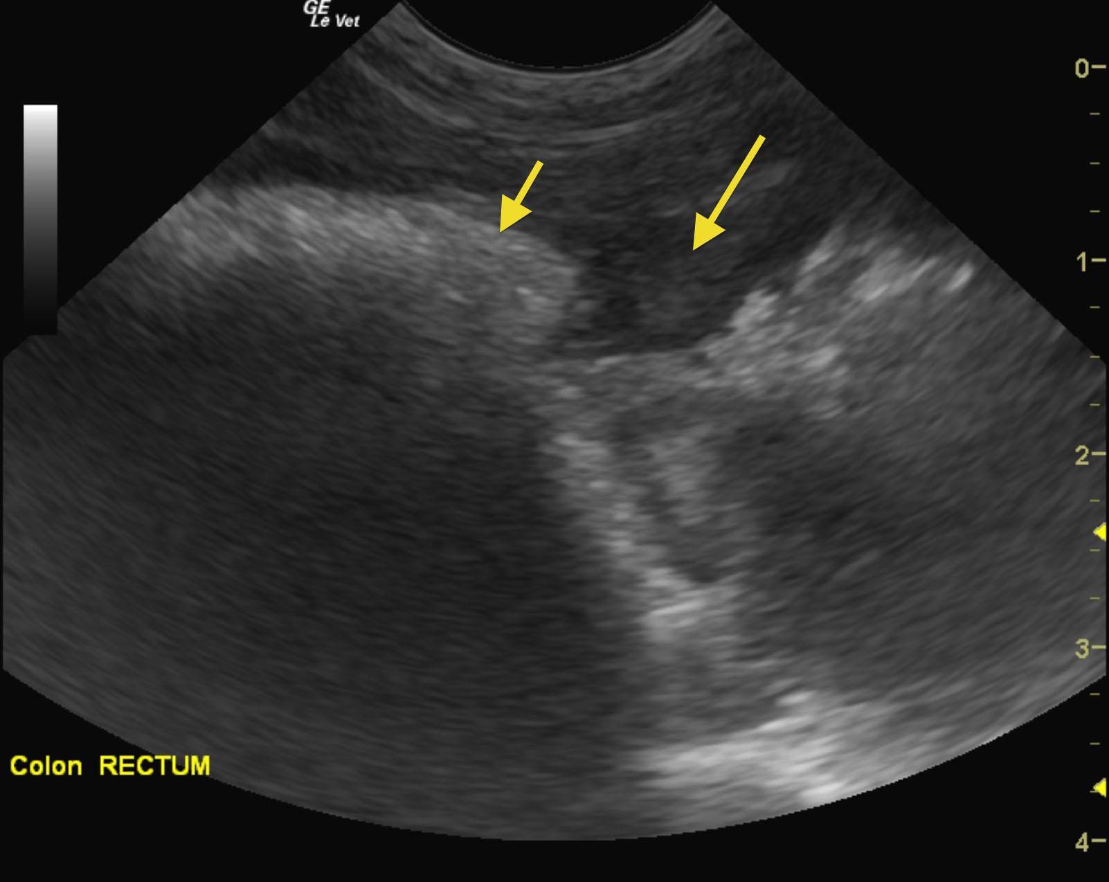
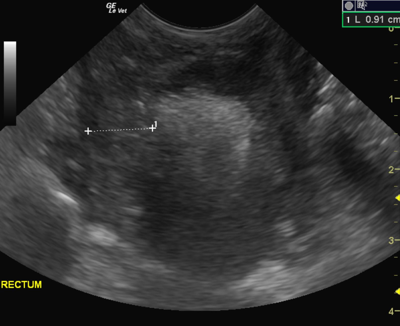
A 6-year-old SF DSH was presented for evaluation of dyschezia because of a rectal stricture that was palpable on rectal palpation. Survey radiographs of the pelvis were within normal limits.
A 6-year-old SF DSH was presented for evaluation of dyschezia because of a rectal stricture that was palpable on rectal palpation. Survey radiographs of the pelvis were within normal limits.