A 1-year-old female Retriever was presented for evaluation of chronic intermittent vomiting, diarrhea, and stunted growth. Abnormalities on serum biochemistry were elevated bile acids, bilirubin, and AST and ALT activity.
A 1-year-old female Retriever was presented for evaluation of chronic intermittent vomiting, diarrhea, and stunted growth. Abnormalities on serum biochemistry were elevated bile acids, bilirubin, and AST and ALT activity.
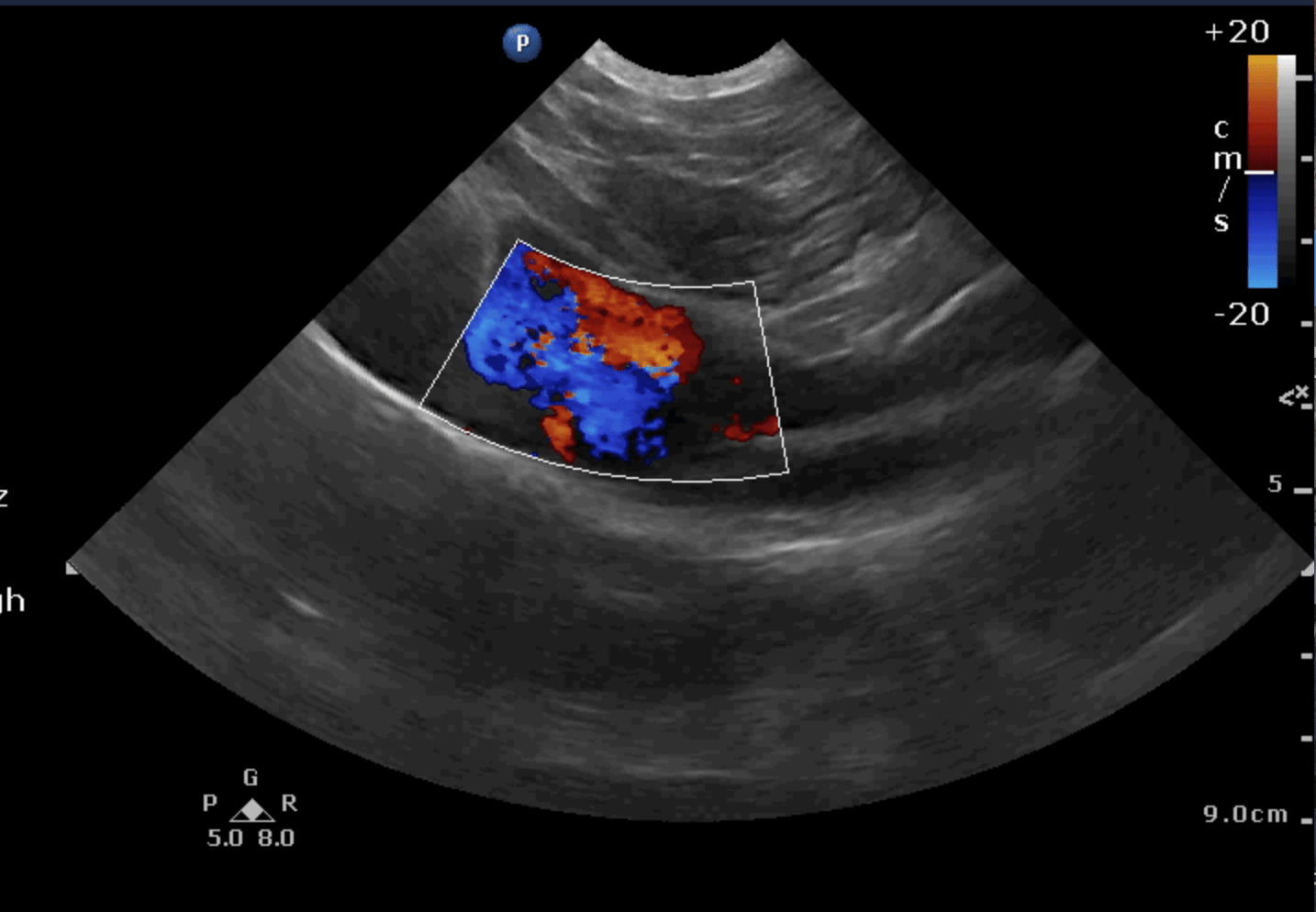
Right divisional intrahepatic shunt.
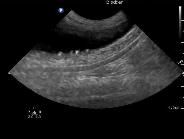
Small bladder calculi.
Recommend referral for intravascular plug placement.
Prognosis is relatively good if interventional therapy can be performed. A traditional surgical
approach could not be performed on this patient given the right divisional position of the shunt.
Vascular plug therapy; however, in my experience has been quite successful with this type of shunt.
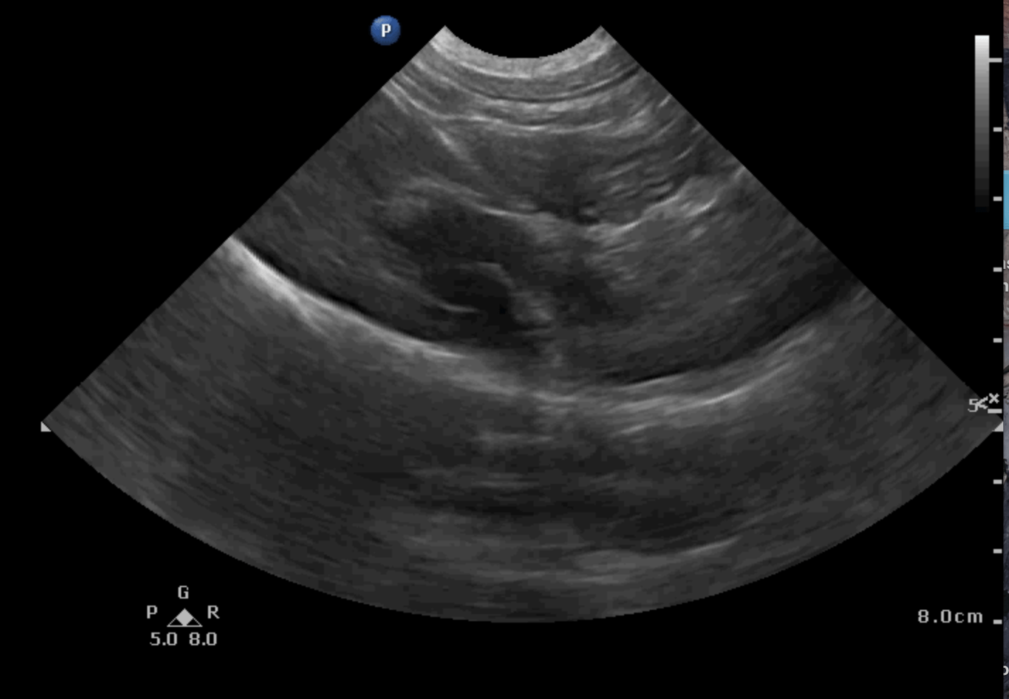
The liver in this patient was subnormal in size with an intrahepatic shunt consistent with right divisional shunt measuring approximately 1 cm in width deriving from the right branch of the portal vein and in intrahepatic fashion entering into the vena cava. This should be amenable to vascular plug therapy with interventional radiology at a referral facility or university. Gallbladder was over distended. Vena cava 1.15 cm. Aorta 1.07 cm. Portal vein 0.95 cm; largely 1:1:1 ratio. No evidence of extrahepatic shunts noted Pelvic urethra was unremarkable imaged 3 cm beyond the cystourethral junction. Small bladder calculi noted. Uterine body was mildly thickened, slight heterogenous changes 0.29 cm. Left ovary uniform 0.96 cm.
None
Porto-systemic shunt, primary portal vein hypoplasia, congenital fibrosis, chronic-active hepatitis, cirrhosis from prior acute hepatic insult
None