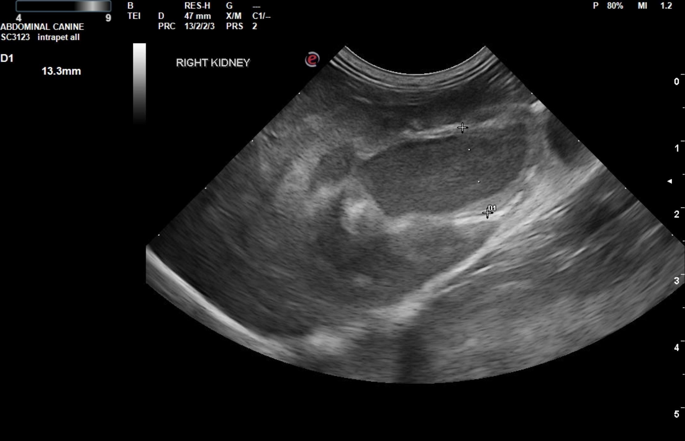
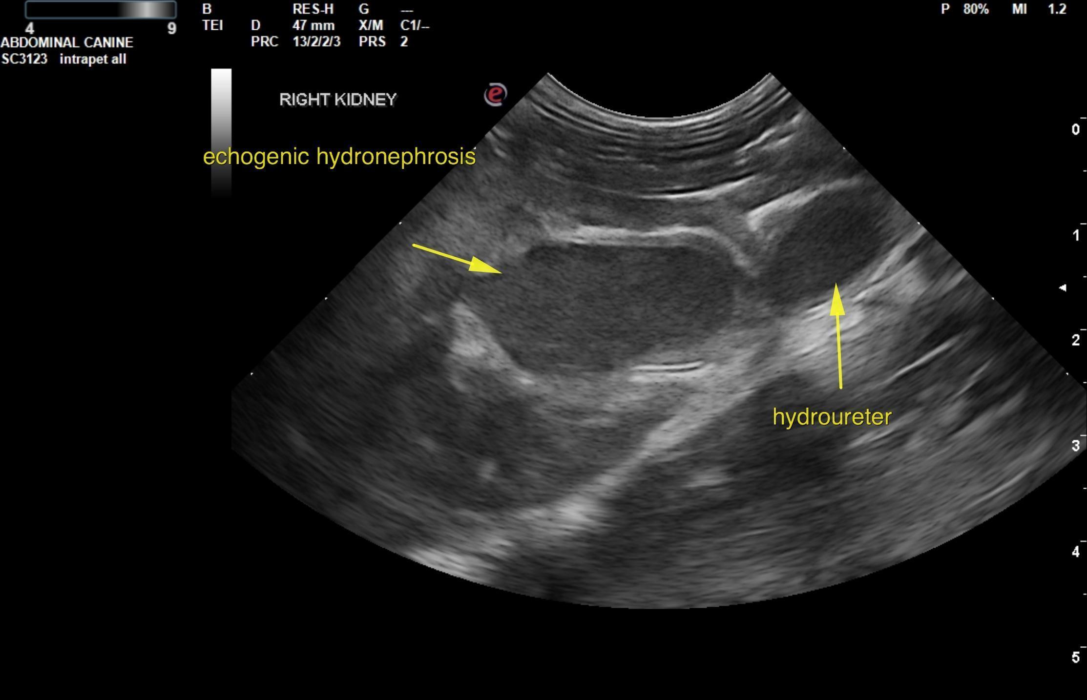
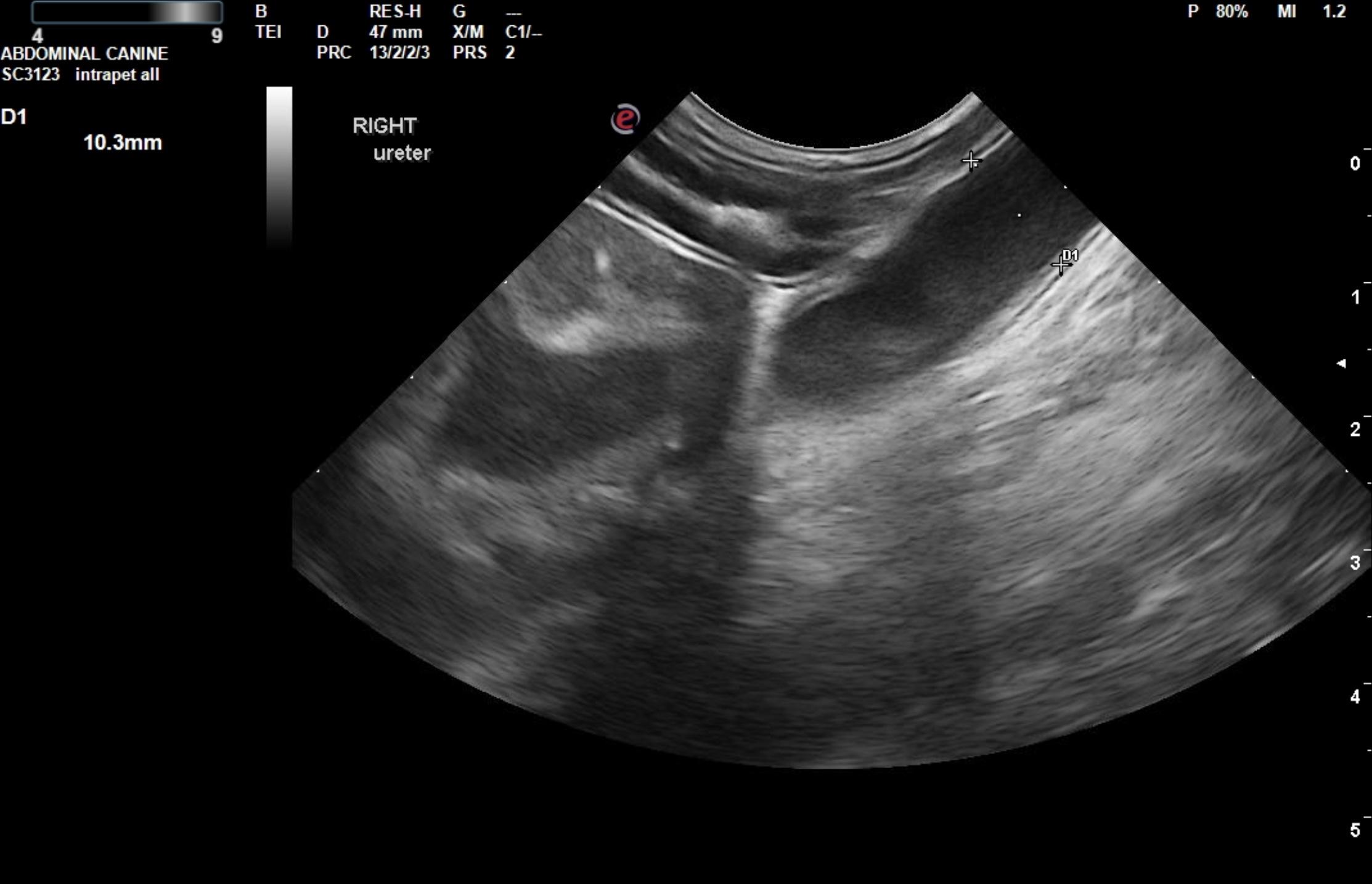
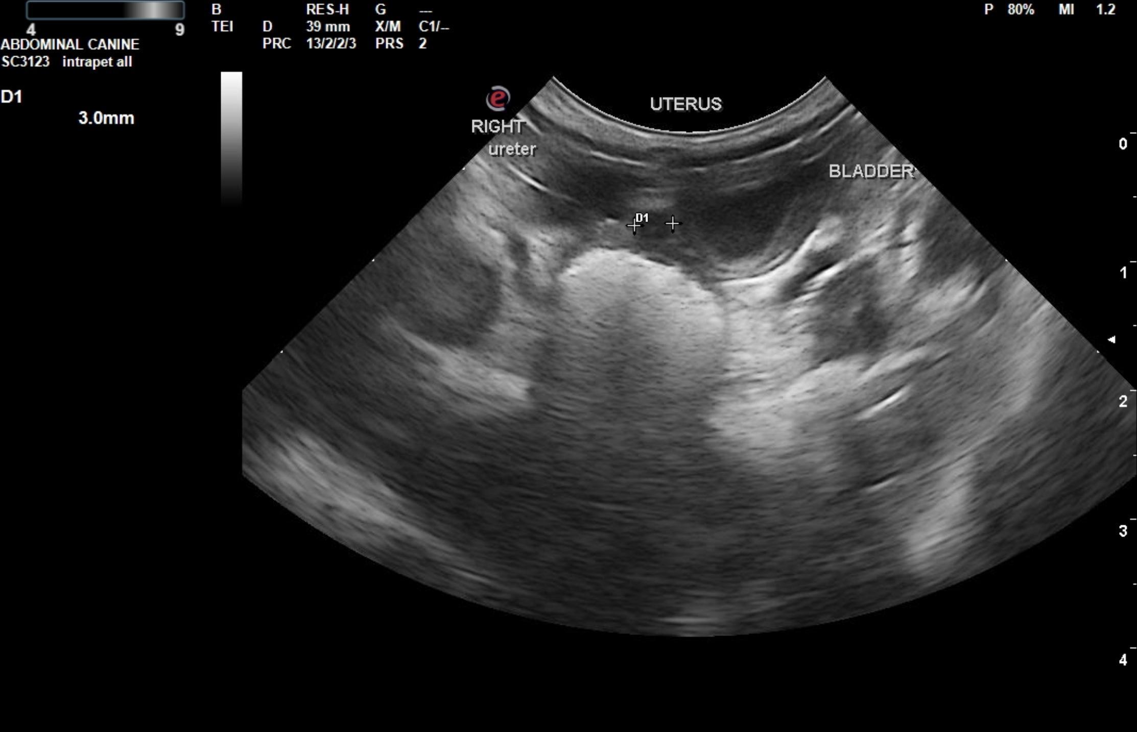
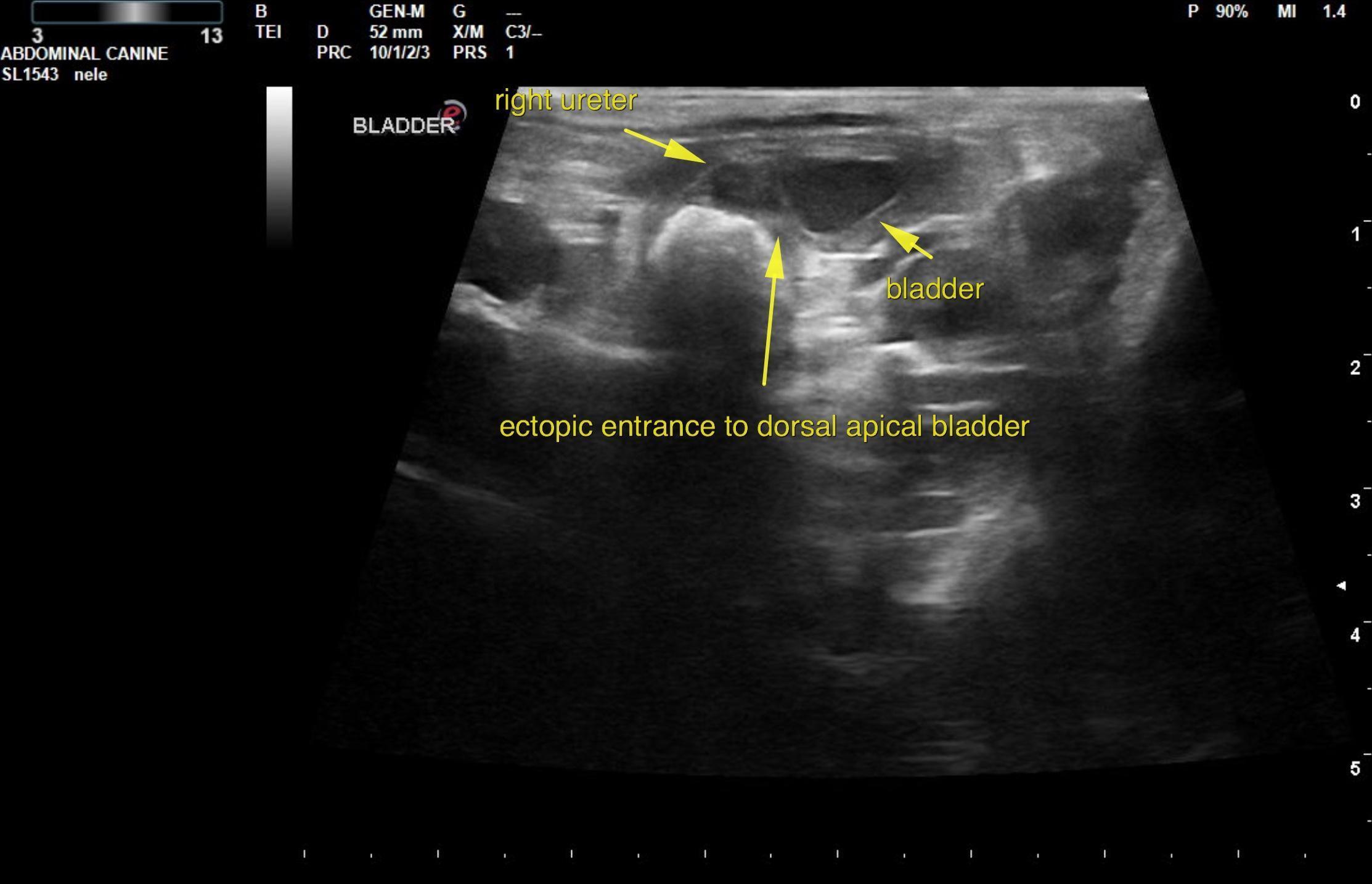
A 3-month-old intact female Golden Doodle was presented for evaluation of UTI and pollakiuria that had improved with antibiotic therapy but relapsed once the course was completed.
A 3-month-old intact female Golden Doodle was presented for evaluation of UTI and pollakiuria that had improved with antibiotic therapy but relapsed once the course was completed.