RAD – Bilateral elbow dysplasia with medial coronoid pathology in a 3 year old MN Labrador Retriever

Case Study

RAD – Bilateral elbow dysplasia with medial coronoid pathology in a 3 year old MN Labrador Retriever

This 3 year old MN Labrador Retriever mix has a history of chronic right fron lameness that is worse after exercise.

This 3 year old MN Labrador Retriever mix has a history of chronic right fron lameness that is worse after exercise.

Image Interpretation

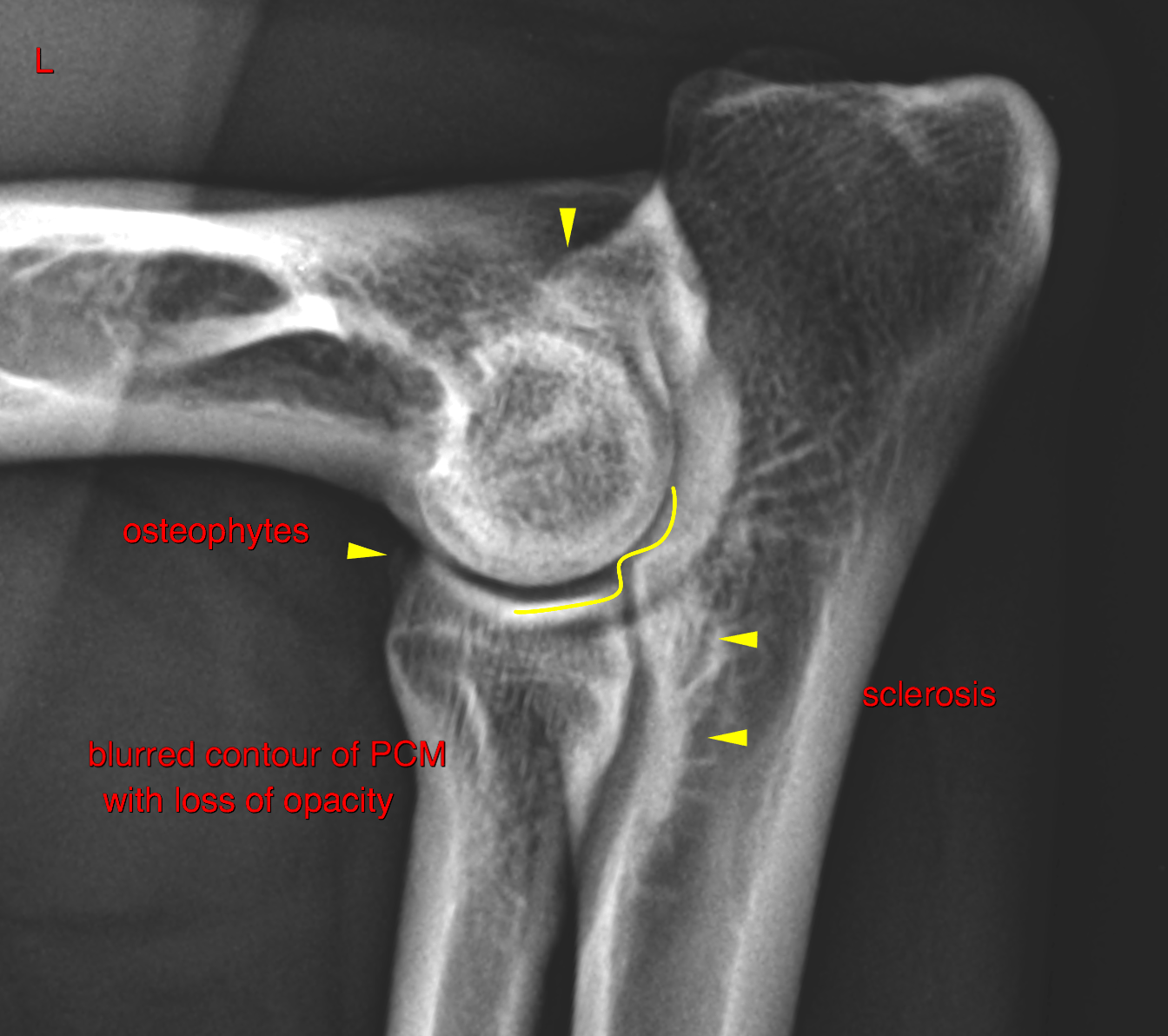
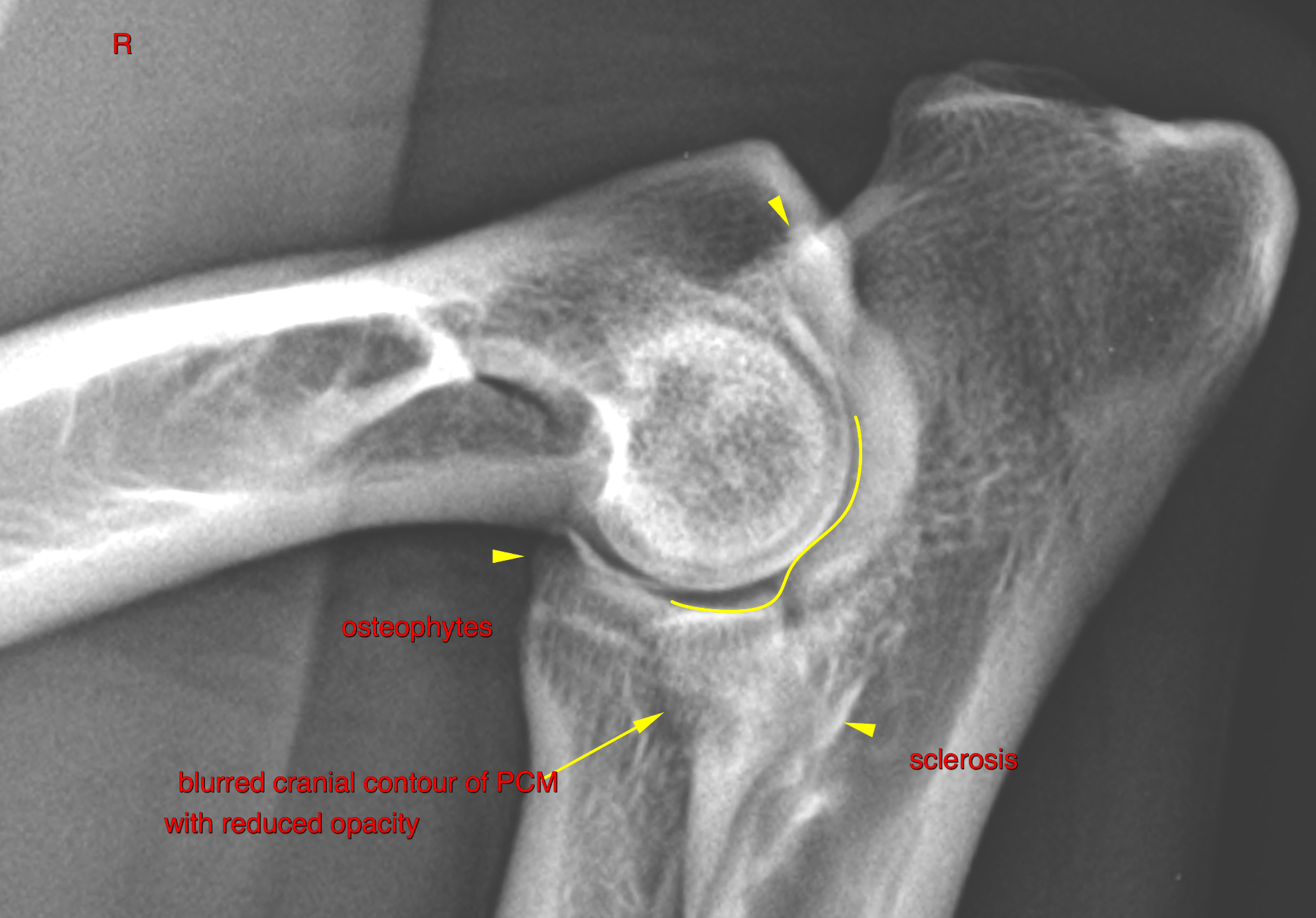
right and left elbows:  The cranial contour of the medial coronoid process (PCM) is blurred and reduced inopacity. Asymmetry of the humeroradial and humeroulnar joint space is noted withmild narrowing of the humeroulnar joint space and a mild radioulnar step formation. A mild amount of osteophytes are noted at the proximocranial aspect of the radius andmproximal surface of the anconeal process as well as sclerosis of the trochlear notch of the ulna.

right and left carpi: A minimal osteophyte formation is noted at the dorsal aspect of the carpometacarpal joint.


DX

Bilateral elbow dysplasia in terms of medial coronoid pathology and mild elbow joint incongruity with mild secondary osteoarthritis

Outcome

CT of both elbows and arthroscopy (arthrotomy) of the right elbow is recommended.
Consider surgical revision of the left elbow joint as well depending on the results of
the CT or in case of clinical lameness of the left front limb.

Patient Information

Patient Name : Ace Culbertson/Golden Isles
Gender : Male, Neutered
Species : Canine
Type of Imaging : Ultrasound
Status : Complete
Liz Wuz Here : Yes
Code : 17_00084

Clinical Signs

  • Lameness

Images

bildschirmfoto_2016-09-24_um_13bildschirmfoto_2016-09-24_um_17

Clinical Signs

  • Lameness
Skip to content