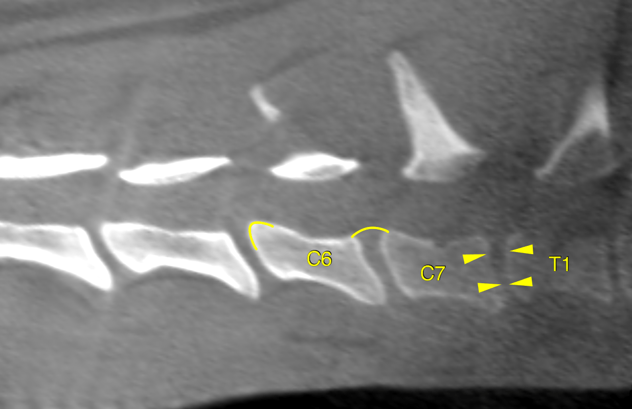
This 3 year old MN Australian Shepherd mix dog presented for apparent pain of 2 months duration. Cries out with sudden movement, ambulates with head parallel to spine. Lays down while eating.
Physical exam: Hopping and CPs normal. No palpable pain
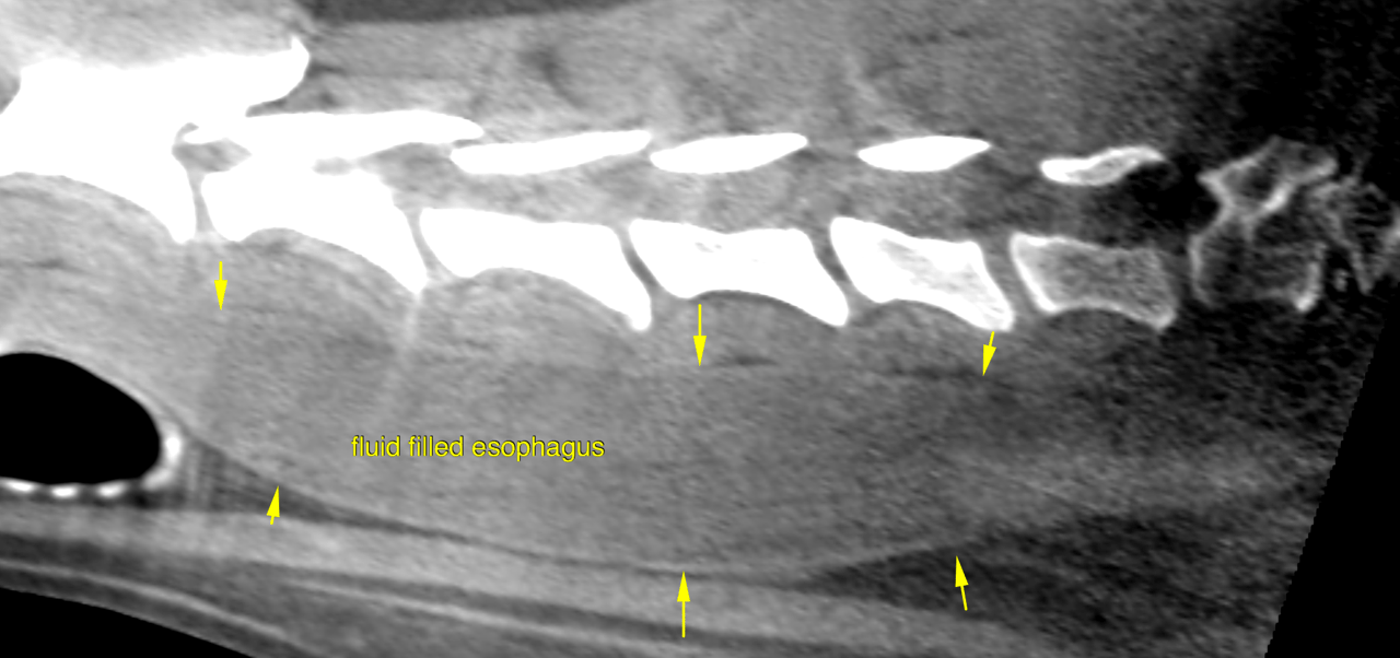
This 3 year old MN Australian Shepherd mix dog presented for apparent pain of 2 months duration. Cries out with sudden movement, ambulates with head parallel to spine. Lays down while eating.
Physical exam: Hopping and CPs normal. No palpable pain