RAD – Age related thoracic changes in an 8 year old FS Boxer dog

Case Study

RAD – Age related thoracic changes in an 8 year old FS Boxer dog

The patient is an 8 year old FS Boxer dog presented with acute hypersalivation of 2 hours duration.  Diagnosed with cutaneous mast cell tumor 2 years ago.

Physical Exam: painful cranial abdominal palpation, hypersalivation

The patient is an 8 year old FS Boxer dog presented with acute hypersalivation of 2 hours duration.  Diagnosed with cutaneous mast cell tumor 2 years ago.

Physical Exam: painful cranial abdominal palpation, hypersalivation

Image Interpretation

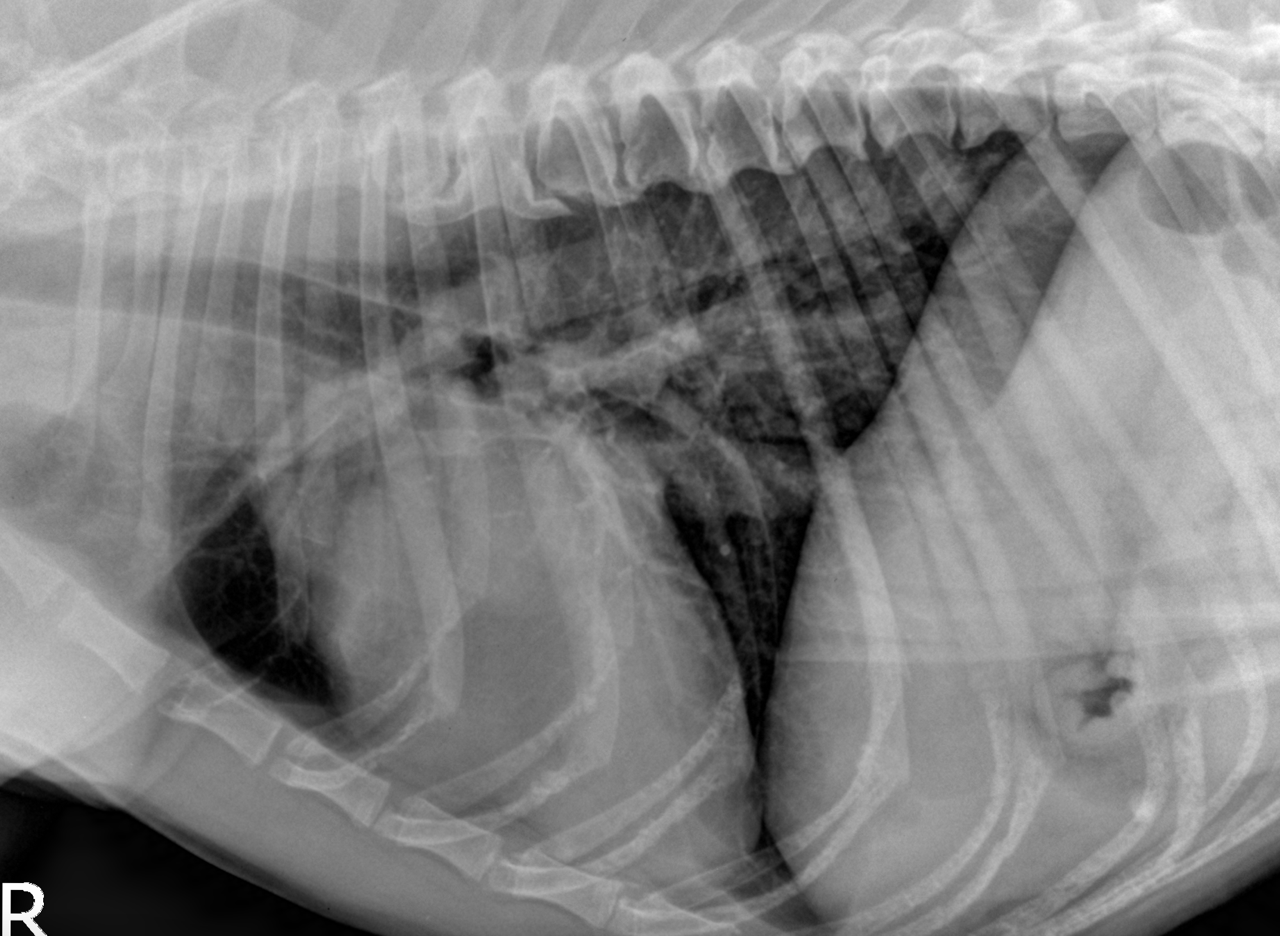
Rads – right lateral and VD view of thorax and abdomen – Osseous structures:
Overall moderate degenerative changes were associated with the axial skeleton
including incomplete bridging spondyloses with ossification of the ventral longitudinal
ligament form T4 to T11. The width of the intervertebral disc spaces is maintained.
There are mild to moderate spondylarthrotic changes. The neighboring endplates of T6
through T9 reveal centrally located well delineated semicircular defects consistent with
Schmorl’s nodes.
One of the shoulder joints reveals mild osteoarthritic changes.
Intrathoracic structures:
The degree of inspiration is moderate.
The trachea, mediastinum and pleural space are within normal limits. The mediastinal
lymph nodes are not seen. The lung reveals a mild generalized bronchointerstitial
pattern. There is no evidence of interstitial nodules.
The cardiac silhouette is within normal limits for size and shape. The major vessels
and pulmonary vasculature are within normal limits. There is no sign of vascular
congestion.
Intraabdominal structures: 
The serosal detail is within normal limits. The liver is contained within the costal arch,
the liver margins are pointed. The spleen, kidneys and urinary bladder are within
normal limits. The stomach is empty except for a mild amount of gas. The small
intestinal loops are non dilated and of even diameter. The colon contains a mild
amount of fecal material.

DX

The radiographic findings of the thorax are compatible with expected age related changes. There is no sign of congestive heart failure or metastatic spread to the lung or mediastinum.

Outcome

Note that some of the common cardiac disorders in Boxers – such as subaortic stenosis
– cannot be ruled out by means of radiographs. Consider an echocardiographic
examination if the clinical findings support cardiac disease.
Disseminated idiopathic sceletal hyperostosis (DISH) and Schmorl’s nodes within the
vertebral endplates – as seen here within the mid thoracic spine – are common findings
in Boxers and usually of no clinical significance

Patient Information

Patient Name : Eden Kelly
Gender : Female, Spayed
Species : Canine
Type of Imaging : Ultrasound
Status : Complete
Liz Wuz Here : Yes

Exam Finding

  • Doughy Abdomen

Images

bildschirmfoto_2016-01-13_um_09
Skip to content