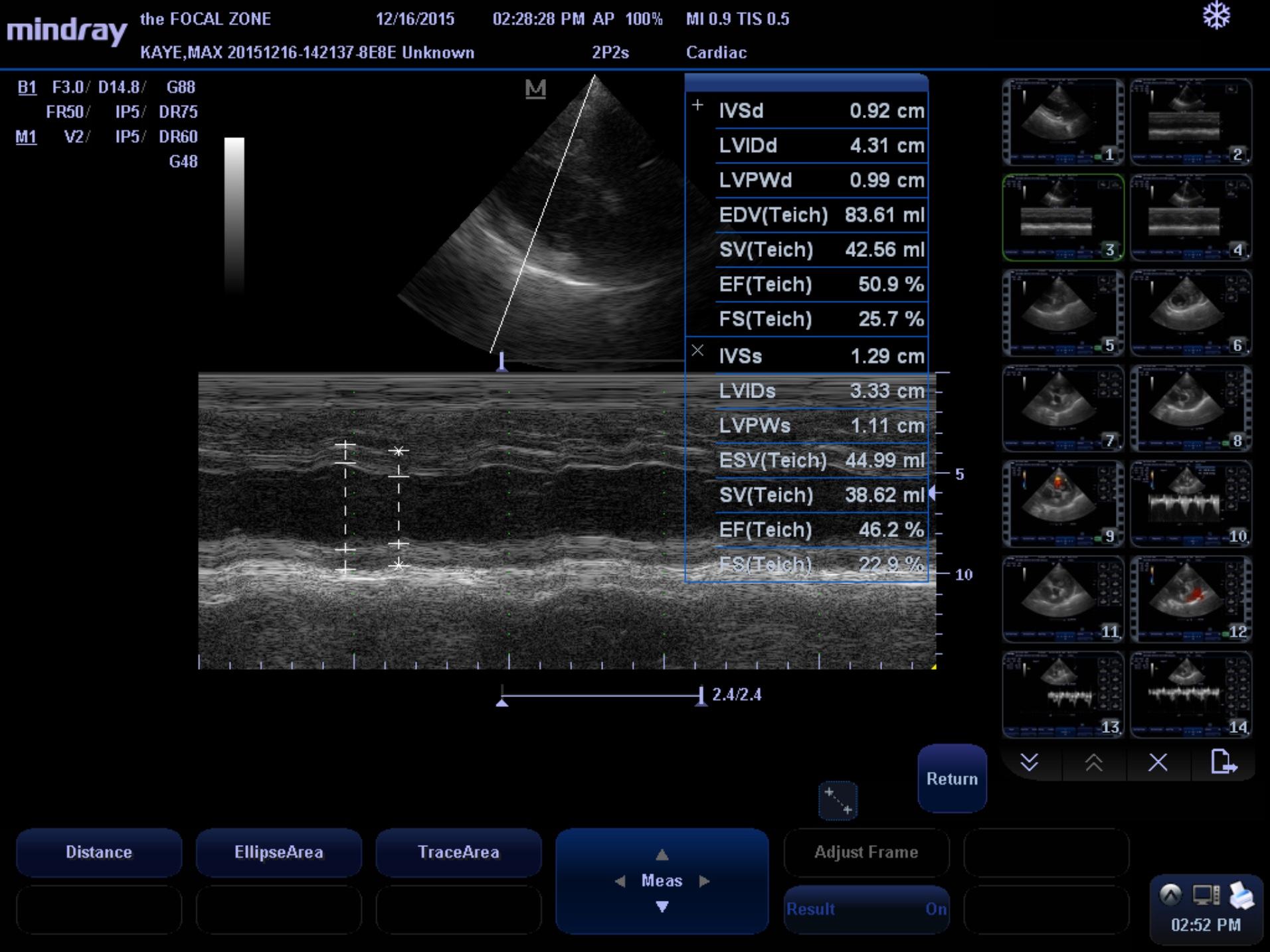
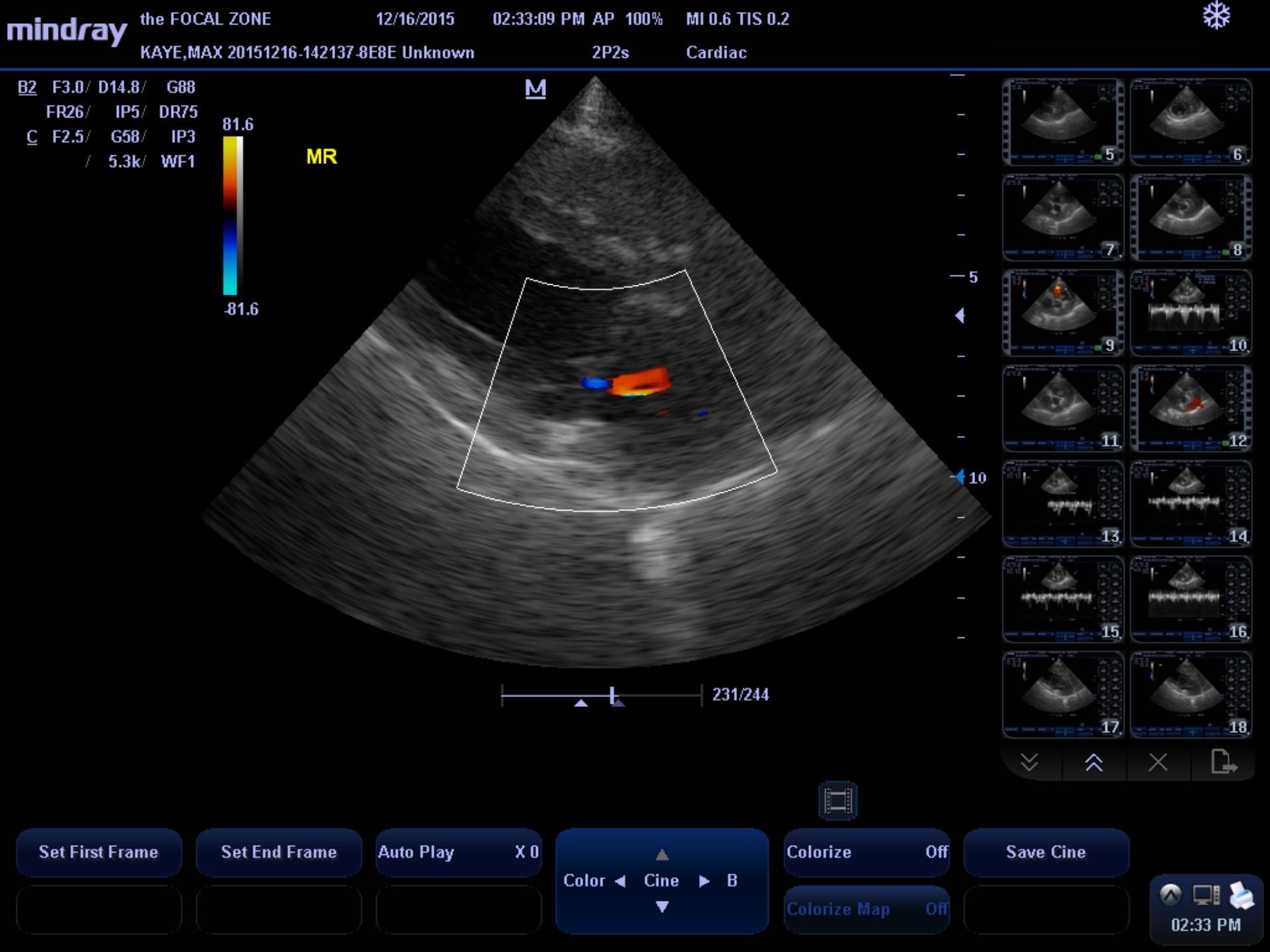
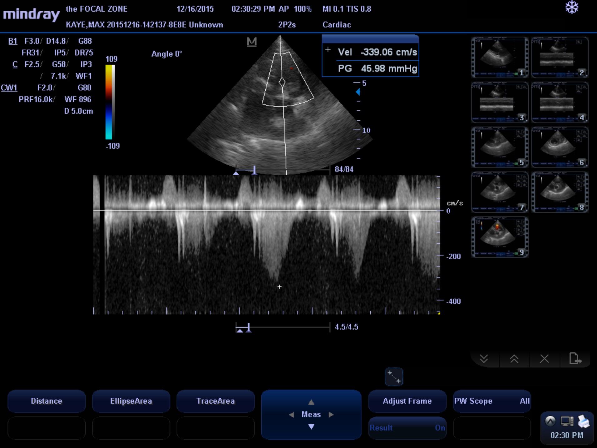
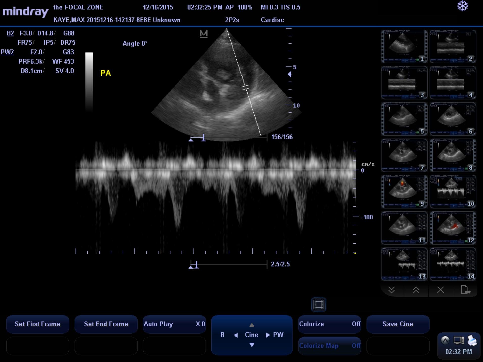
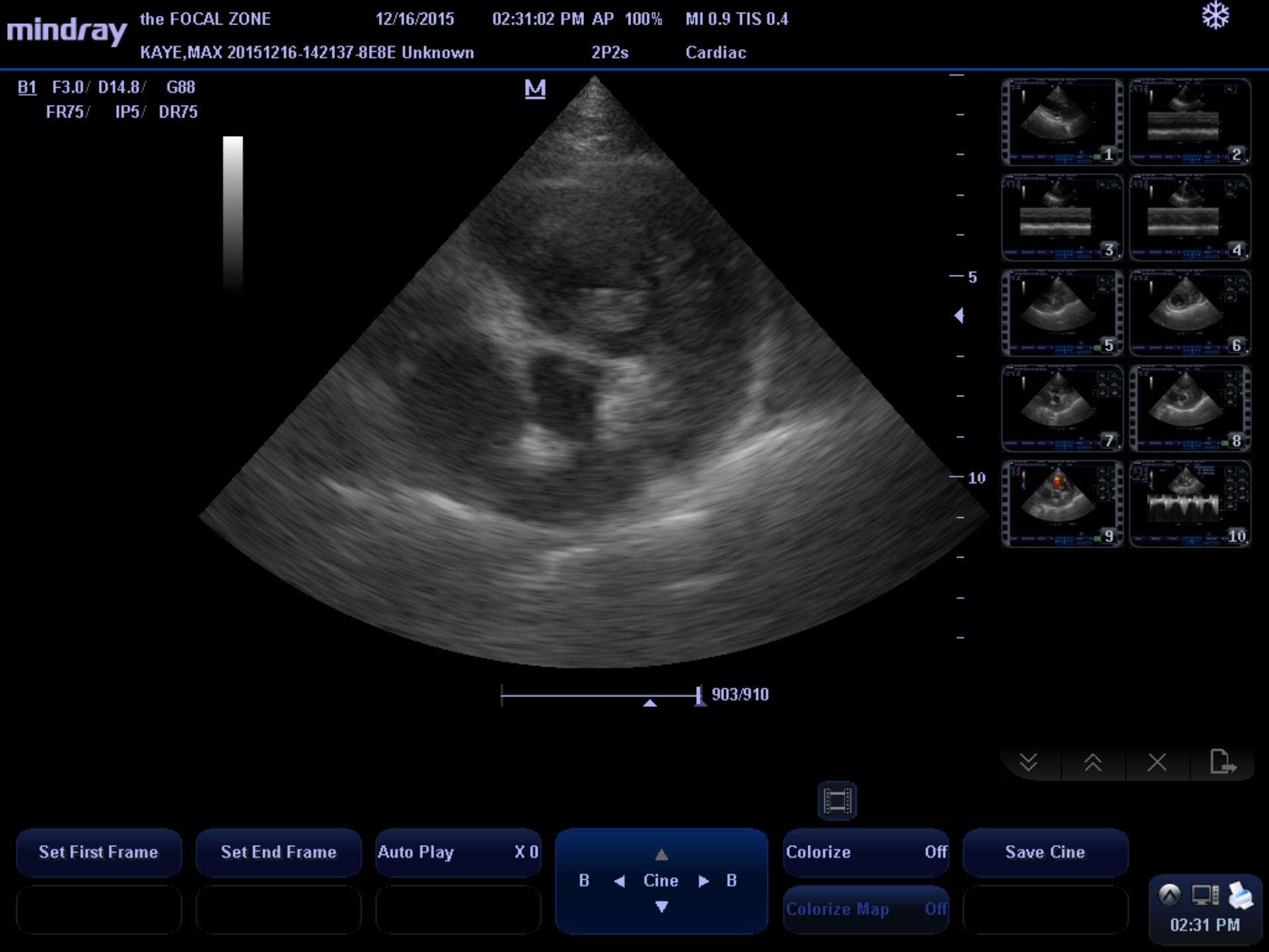
Patient is an 8 year old MN Golden Retriever dog who presented with heavy breathing/panting for the last 2-3 days. Owner has noticed a “pounding heart” when the dog goes out or eats. Physical exam – HR 180, bilateral heart murmur
Chest rads show globoid heart
Patient is an 8 year old MN Golden Retriever dog who presented with heavy breathing/panting for the last 2-3 days. Owner has noticed a “pounding heart” when the dog goes out or eats. Physical exam – HR 180, bilateral heart murmur
Chest rads show globoid heart