13_00038 Grady K Unilateral retinal detachment

Case Study

13_00038 Grady K Unilateral retinal detachment

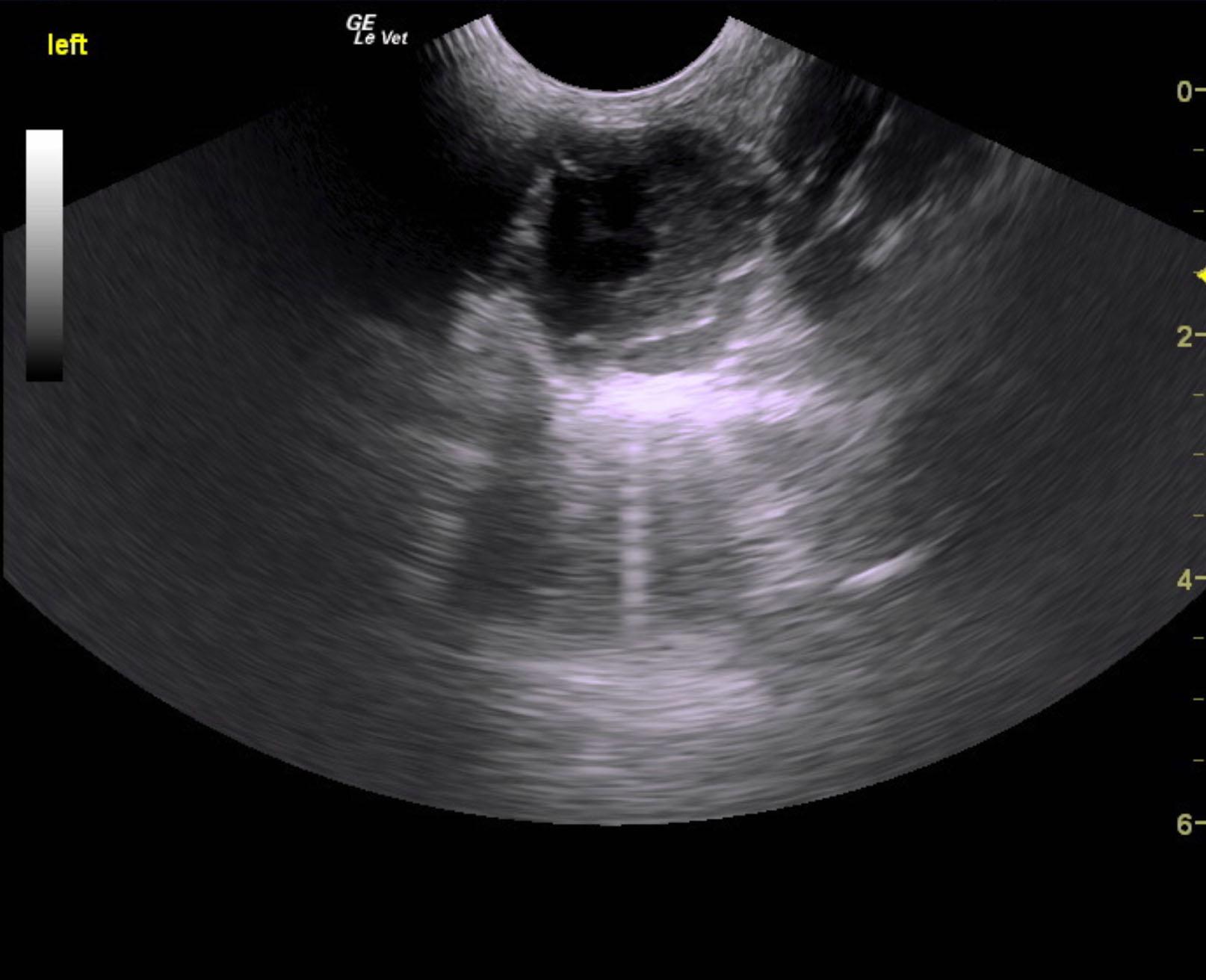
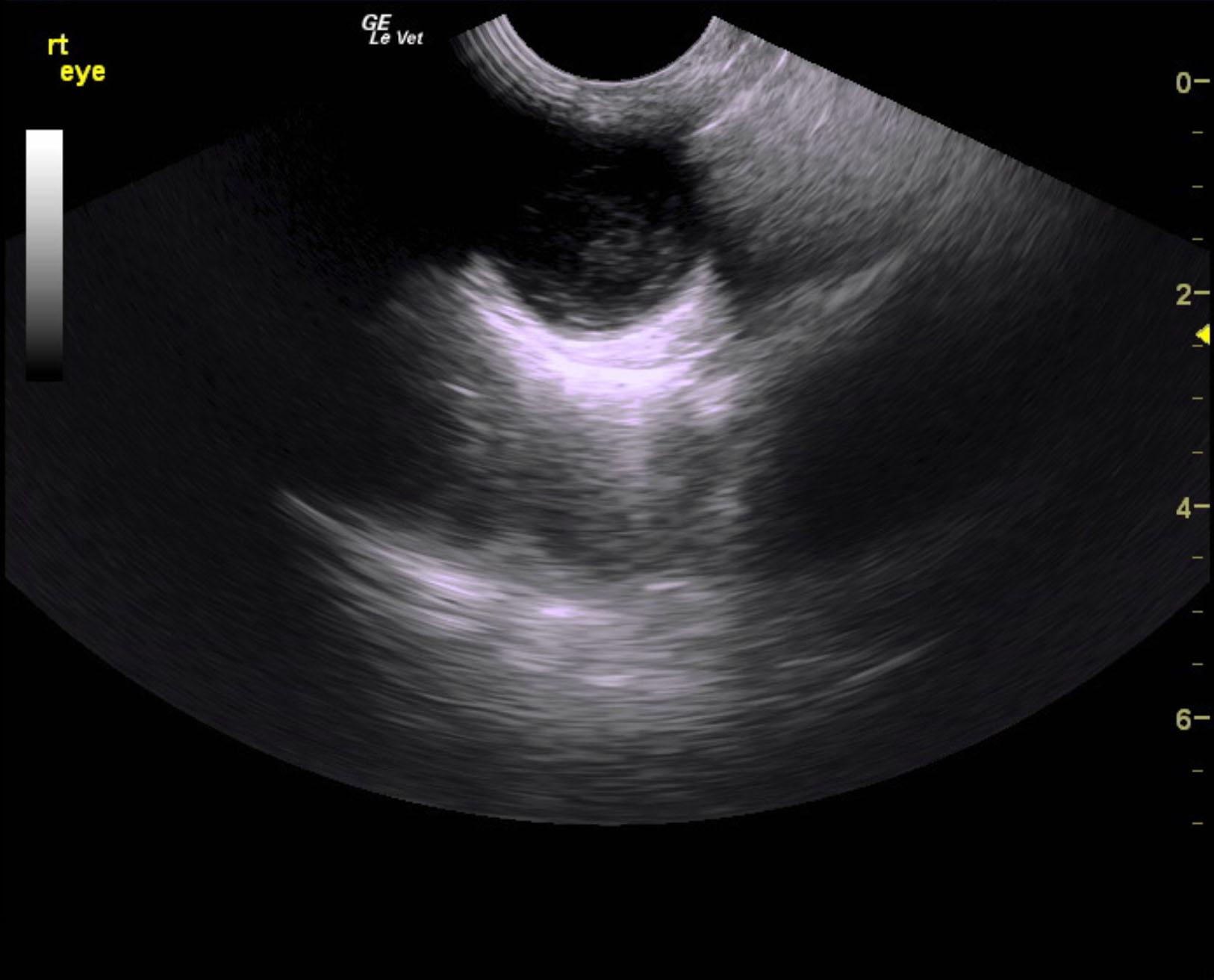
An 11-year-old NM Schnauzer was presented for evaluation of a possible eye tumor and splenic mass. On physical examination, left ocular pressure was increased. CBC showed mild anemia and on survey radiographs possible splenic mass and bladder stones were evident.


Sonographic Differential Diagnosis

Left retinal detachment with posterior chamber debris and posterior uveitis pattern. Confirmation with ophthalmology exam is recommended. Minor posterior uveitis or inflammatory pattern of the right eye. Retina appears intact at the moment.
Systemic blood pressures are recommended. Ophthalmology exam is recommended with medical therapy based on the presentation and ocular pressures.

Image Interpretation

Left eye presented echogenic suspended debris in the posterior chamber with a seagull sign consistent with retinal detachment. Retrobulbar space appeared unremarkable. The right eye revealed minor suspended debris with minor thickened globe consistent with chronic inflammation. Posterior uveitis is suspected. No evidence of neoplasia. The retina appears to intact on the right.

DX

Unilateral retinal detachment

Outcome

None

Clinical Differential Diagnosis

Eye – neoplasia, granuloma, retrobulbar abscess, glaucoma
Spleen – neoplasia, hematoma, granuloma, abscess, infarction

Sampling

None

Patient Information

Gender : Male, Neutered
Species : Canine
Type of Imaging : Ultrasound
Status : Complete

Exam Finding

  • Intraocular pressure increased

Images

grady_krebs_uveitis_retinal_detachment_gluacoma_on_left_eyegrady_krebs_uveitis_retinal_detachment_gluacoma_on_left

CBC

  • RBC, Low
Skip to content