A 9-year-old SF Beagle mix was presented for evaluation of vomiting. Abnormities on CBC and serum biochemistry were hemoconcentration, neutrophilia, elevated bilirubin and cholesterol, hyperglobulinemia, and severely elevated liver enzyme activity.
A 9-year-old SF Beagle mix was presented for evaluation of vomiting. Abnormities on CBC and serum biochemistry were hemoconcentration, neutrophilia, elevated bilirubin and cholesterol, hyperglobulinemia, and severely elevated liver enzyme activity.
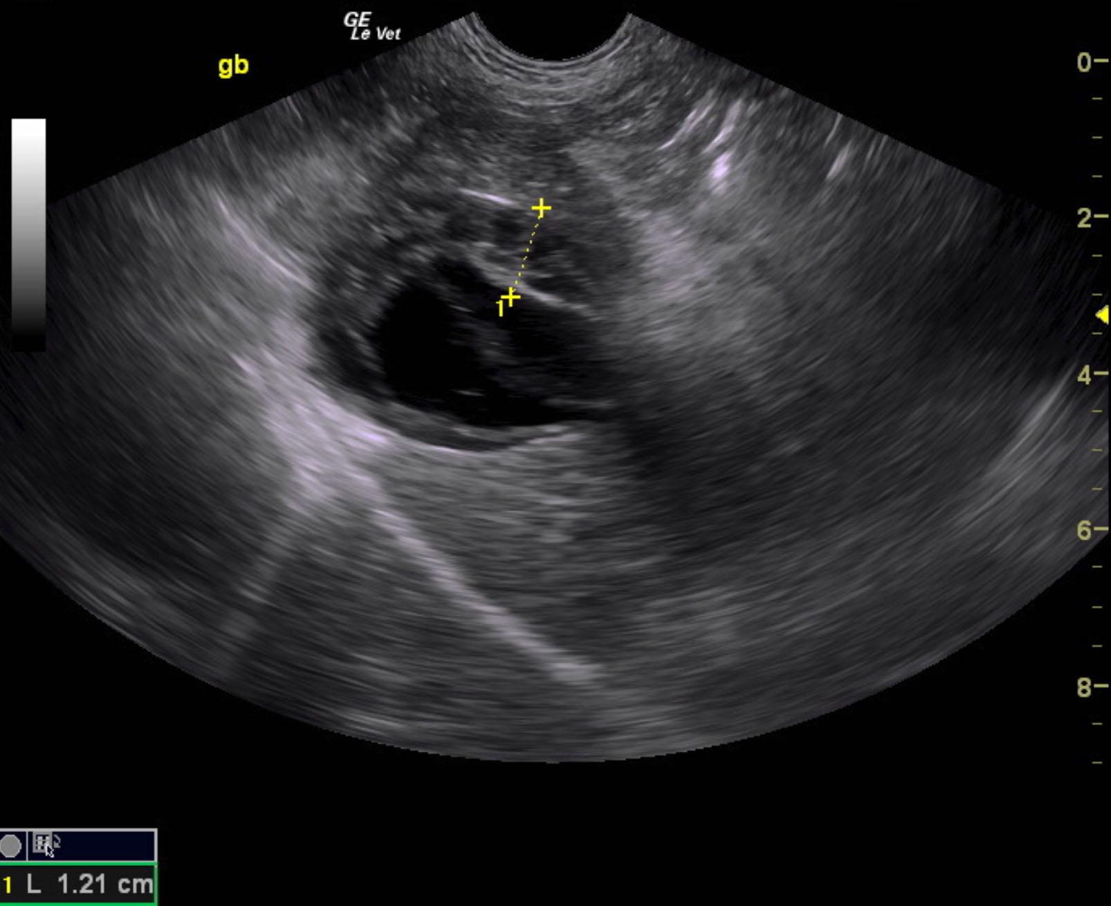
Inflamed gallbladder with possible history of collapse. Potential history of perforation and adhesions. Concurrent cholangiohepatitis pattern. Recommend immediate cholecystectomy and common bile duct lavage. Some aspect of mucocele are noted; however, this may have collapsed and leaked. Regardless the gallbladder is of the primary pathology given the sonographic features most consistent with a combination presentation of aggressive cholecystitis with regional adhesions as well as some aspects of mucocele.
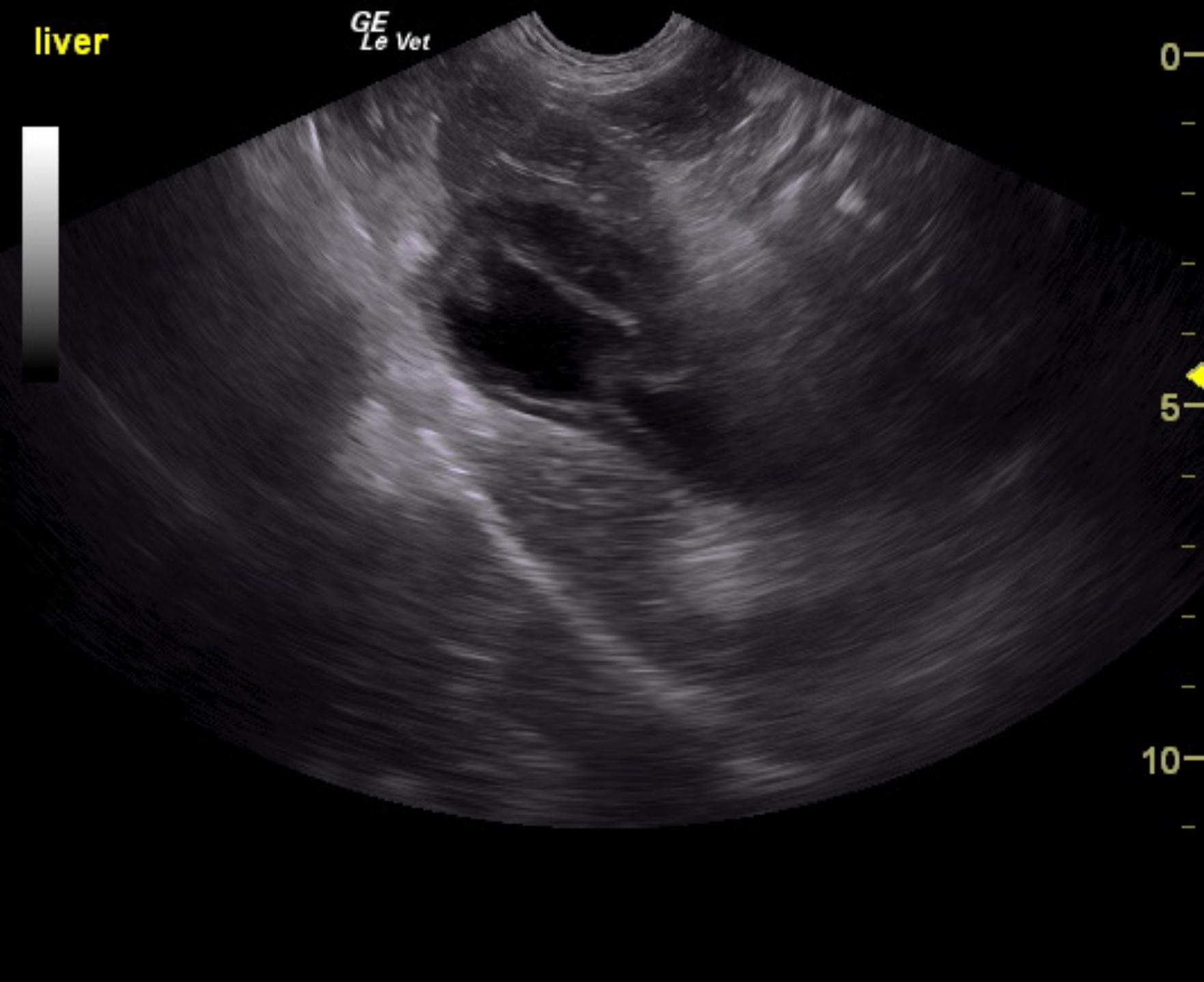
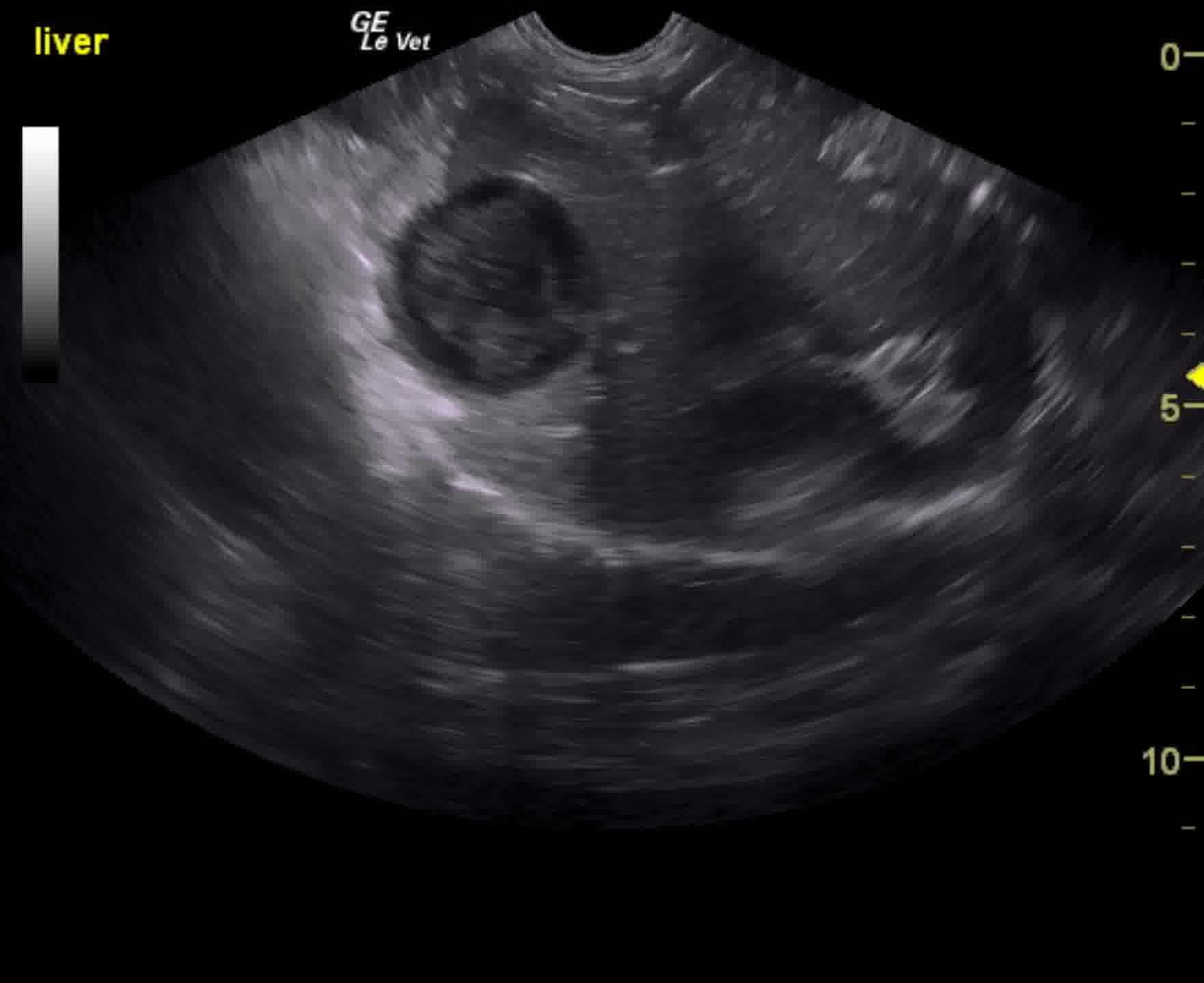
The liver images from right and left intercostal as well as subcostal views revealed subjectively normal liver size, contour, and structure. Parenchymal echogenicity was naturally coarse and hypoechoic to the spleen. Vascular and biliary tracts were of normal volume and no evidence of congestion was noted. Gallbladder wall in this patient was double-layered measuring 1.21 cm. Striating bile and overdistention is noted with adhesion pattern along the neck and the apex. The gallbladder itself measured approximately 4 cm. Increased portal markings noted. Trace amount of free fluid noted around the gallbladder itself. Common bile duct did not appear pathological.
None
Liver – acute hepatitis (viral, bacterial, toxins), neoplasia, abscessation
Gall bladder – cholecystitis, mucocele, obstruction (lith, neoplasia, duodenal/pancreatic disease)
Pancreas – chronic pancreatitis, neoplasia
None