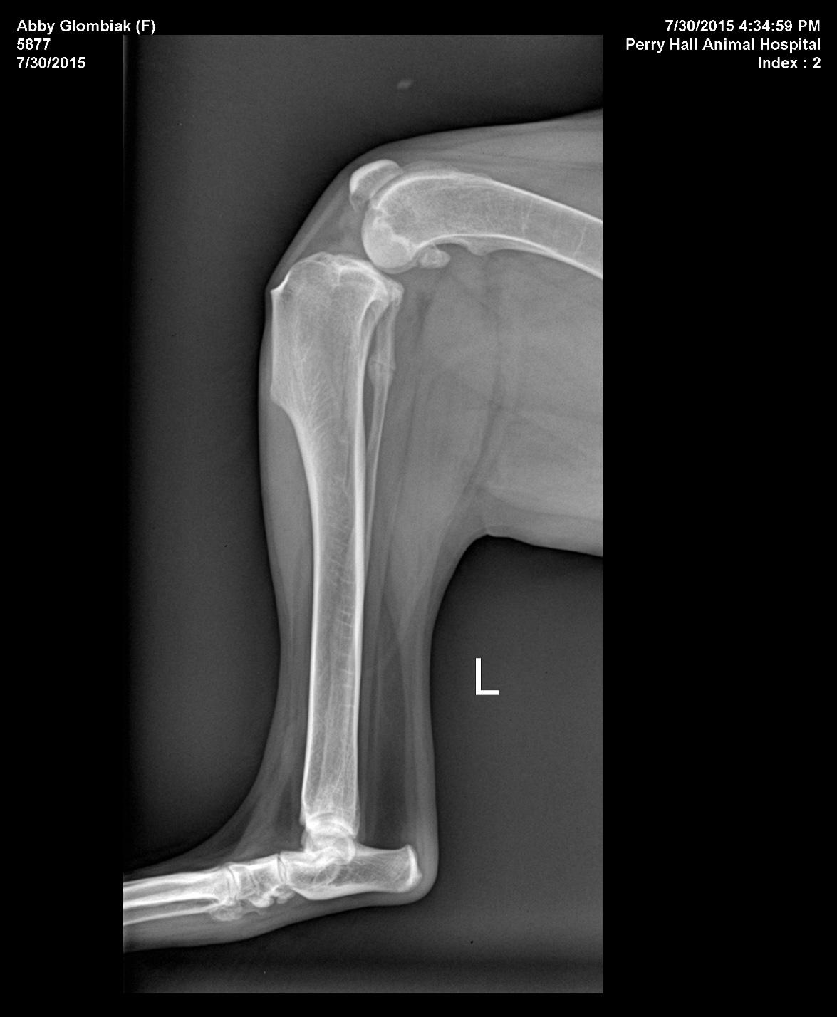
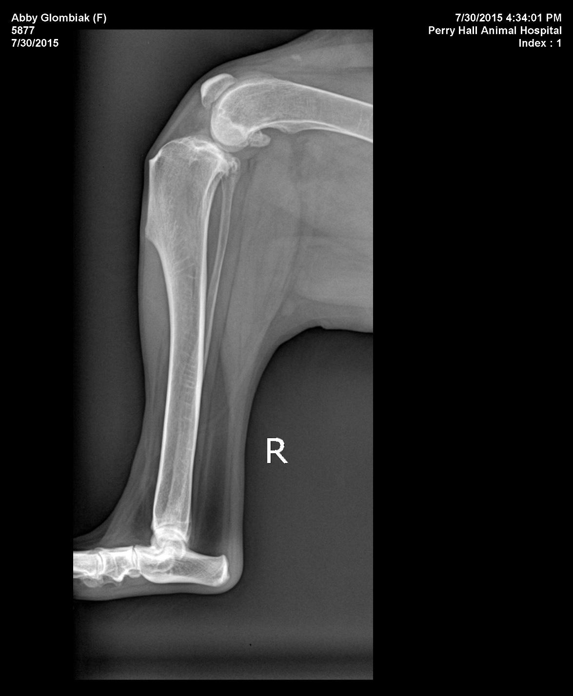
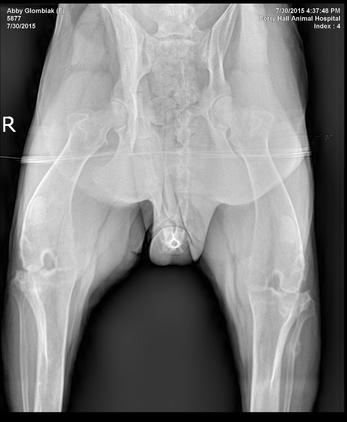
History of left hind leg lameness since February 2015. The patient had a hemilaminectomy on 5/28/5 which alleviated some of the lameness issues initially, but it progressed and the lameness has continued. Physical exam showed partial weight bearing consistently on the left hind leg with positive drawer and cranial tibial thrust. BCS 6.5/9. Neurological exam was WNL.
History of left hind leg lameness since February 2015. The patient had a hemilaminectomy on 5/28/5 which alleviated some of the lameness issues initially, but it progressed and the lameness has continued. Physical exam showed partial weight bearing consistently on the left hind leg with positive drawer and cranial tibial thrust. BCS 6.5/9. Neurological exam was WNL.