The patient is a canine Maltese, SF, 3 years old who was presented for seizures which started 1.5 years ago but are sporadic in nature. Chemistry showed BUN 2.8, low TP 5.3, Albumin 2.3, AST 73, Bile Acids random 125. CBC is wnl.
The patient is a canine Maltese, SF, 3 years old who was presented for seizures which started 1.5 years ago but are sporadic in nature. Chemistry showed BUN 2.8, low TP 5.3, Albumin 2.3, AST 73, Bile Acids random 125. CBC is wnl.
Intrahepatic portosystemic shunt.
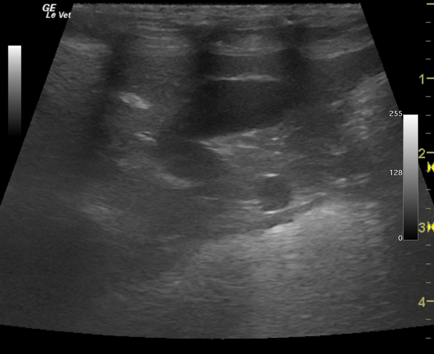
The urinary bladder presented calculus at 0.6 cm and other small calculi that measured 0.2-0.4 cm. The bladder wall was unremarkable.
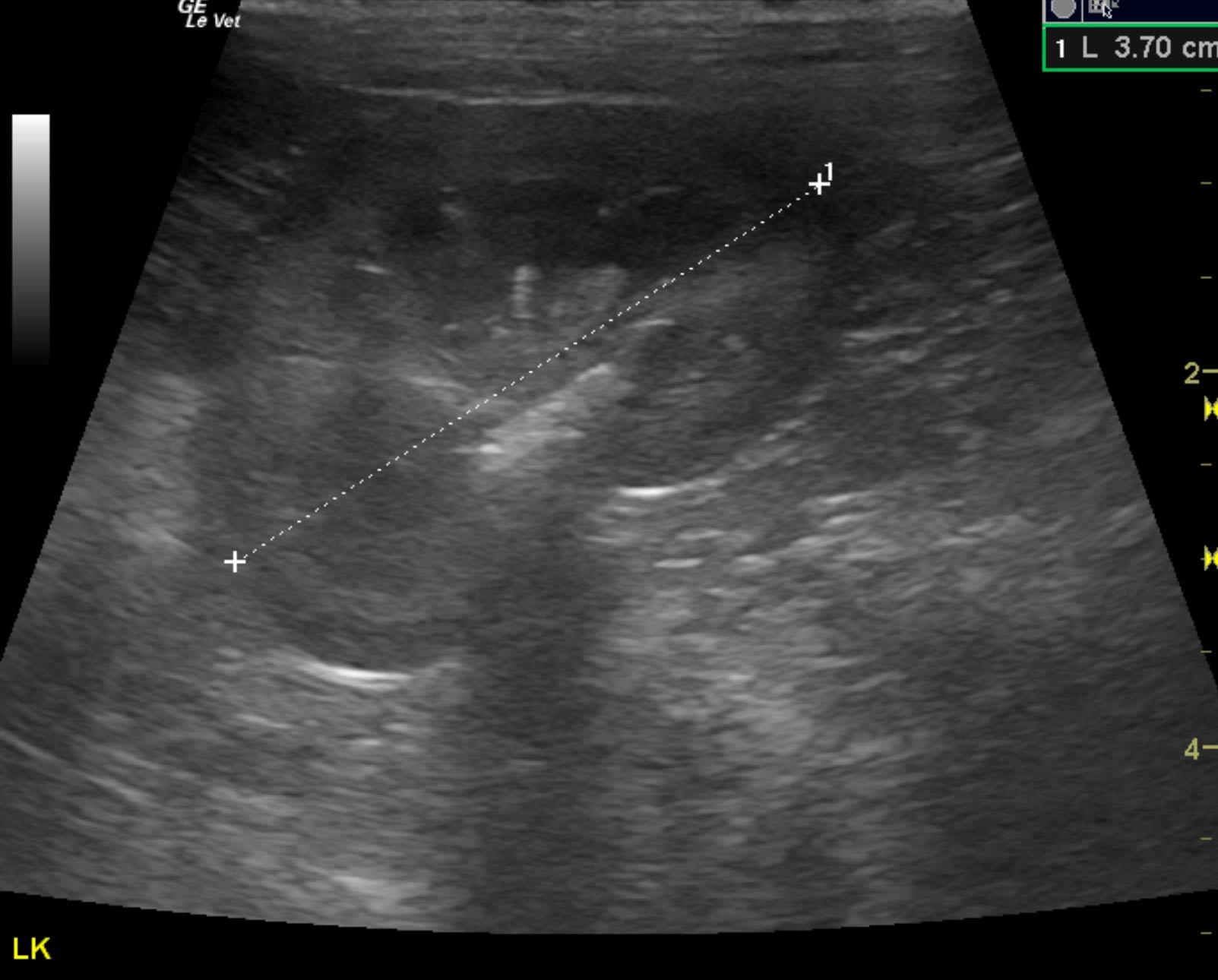
The right kidney was subnormal in size and measured 3.16 cm with cortical remodeling and slight mineralization. The right kidney revealed slight pyelectasia. The patient is likely passing calculi periodically. The left kidney was normal in size at 3.7 cm with dystrophic mineralization and ill defined pelvic fat.
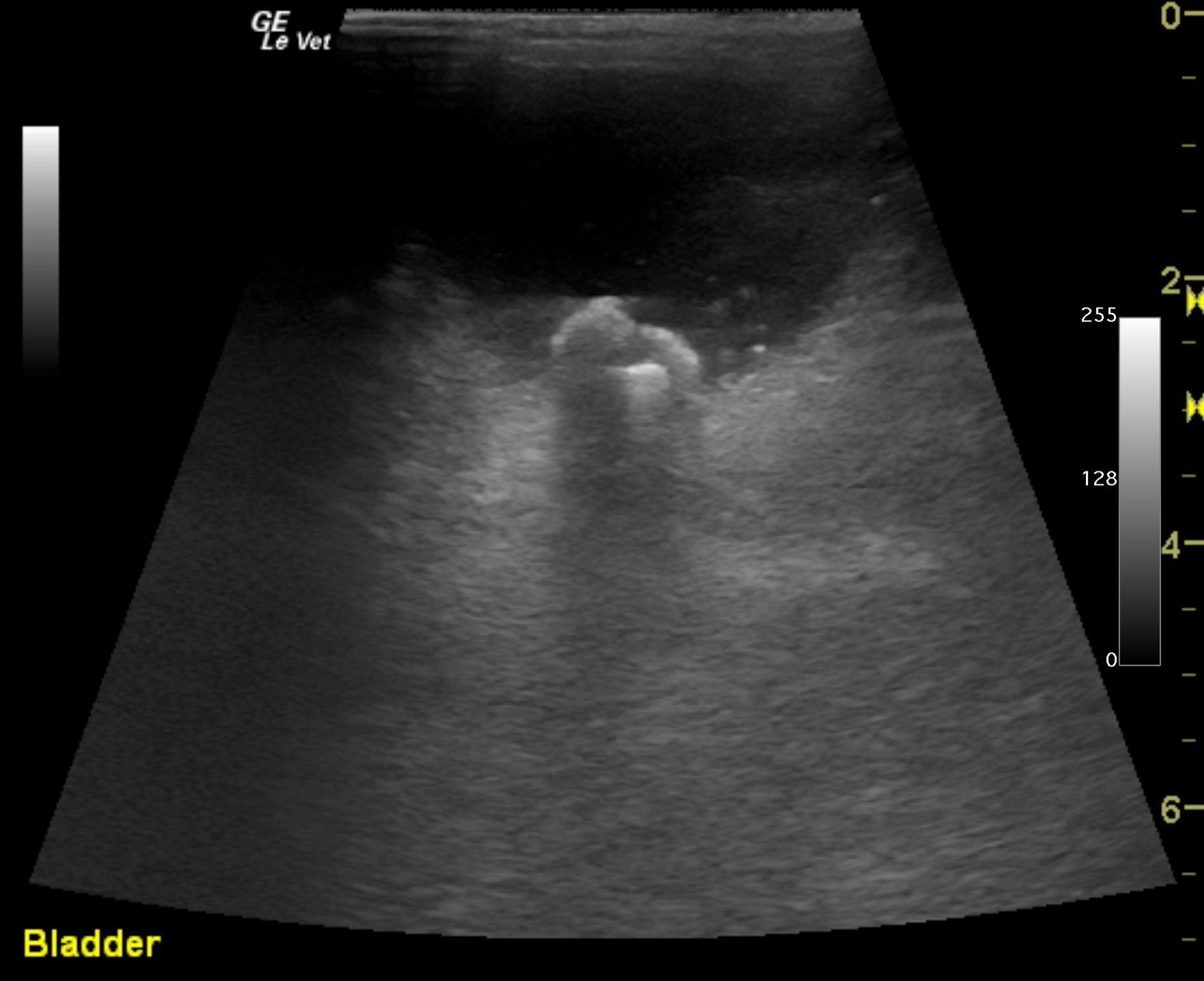
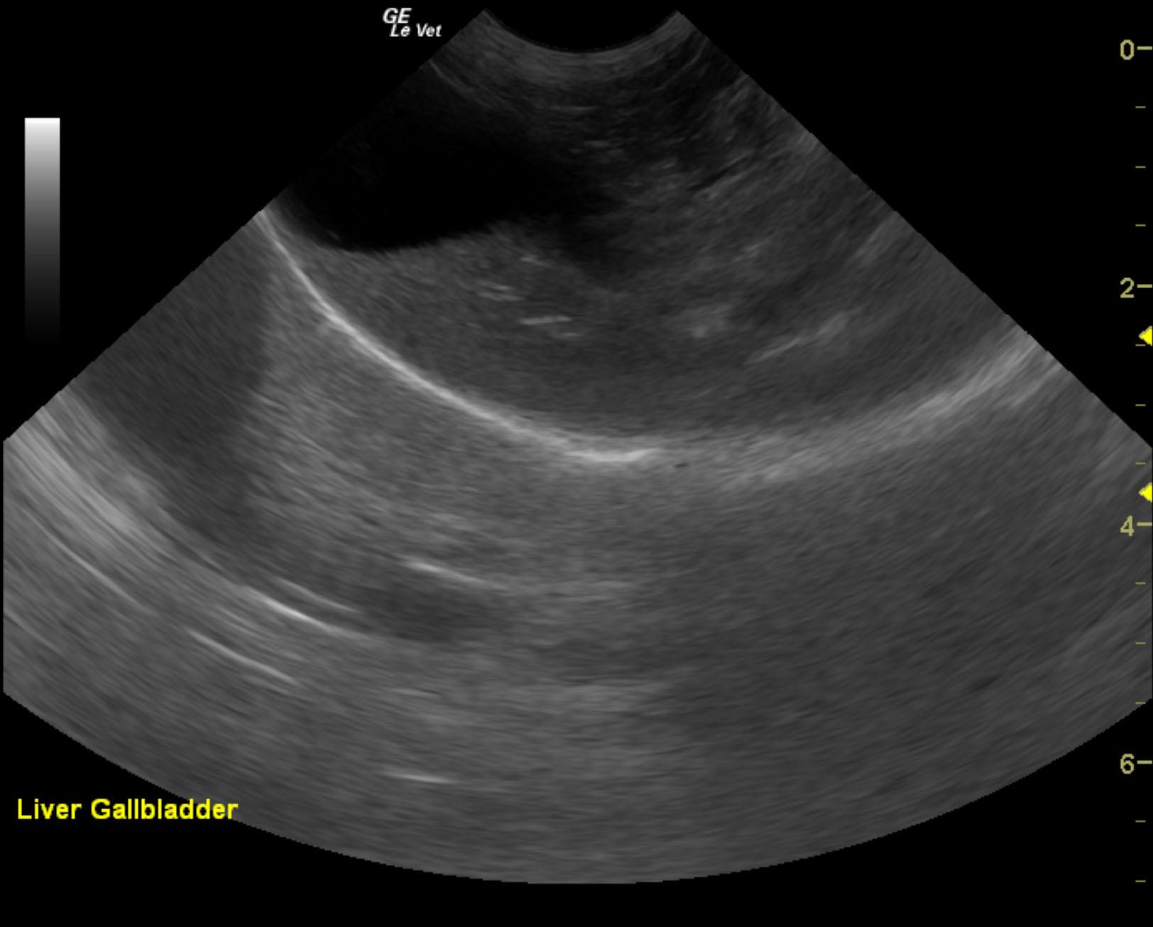
The liver was subnormal in size and coarse in architecture. The portal vein revealed a right divisional intrahepatic shunt connecting to the vena cava within the liver. This is not a surgical type of shunt; however, vascular plug placement with interventional radiology would be ideal in this case. Even though this is a small breed this is an intrahepatic shunt and not extrahepatic. The intrahepatic branch of the shunt measured approximately 0.4 cm at the division from the portal vein and 0.8 cm at the level of the vena cava. However, the prehepatic portal vein, vena cava and aorta were 1:1:1, which is what we would expect for an intrahepatic shunt.
None
None