A 3-year-old NM DSH was presented for evaluation of ADR and seizure activity. On physical examination, the patient was unkempt. Urinalysis was normal and the only change on blood work was subnormal urea.
A 3-year-old NM DSH was presented for evaluation of ADR and seizure activity. On physical examination, the patient was unkempt. Urinalysis was normal and the only change on blood work was subnormal urea.
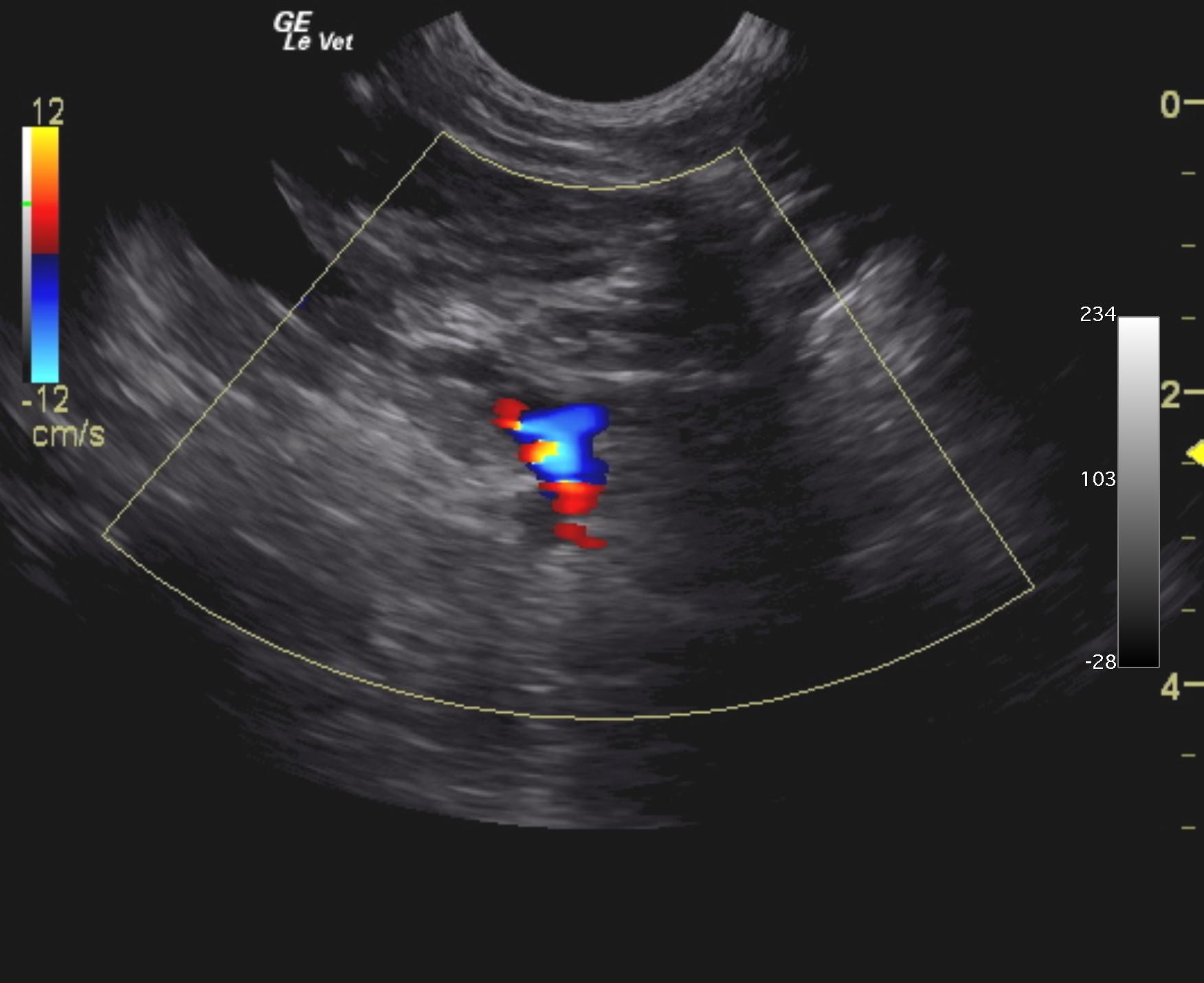
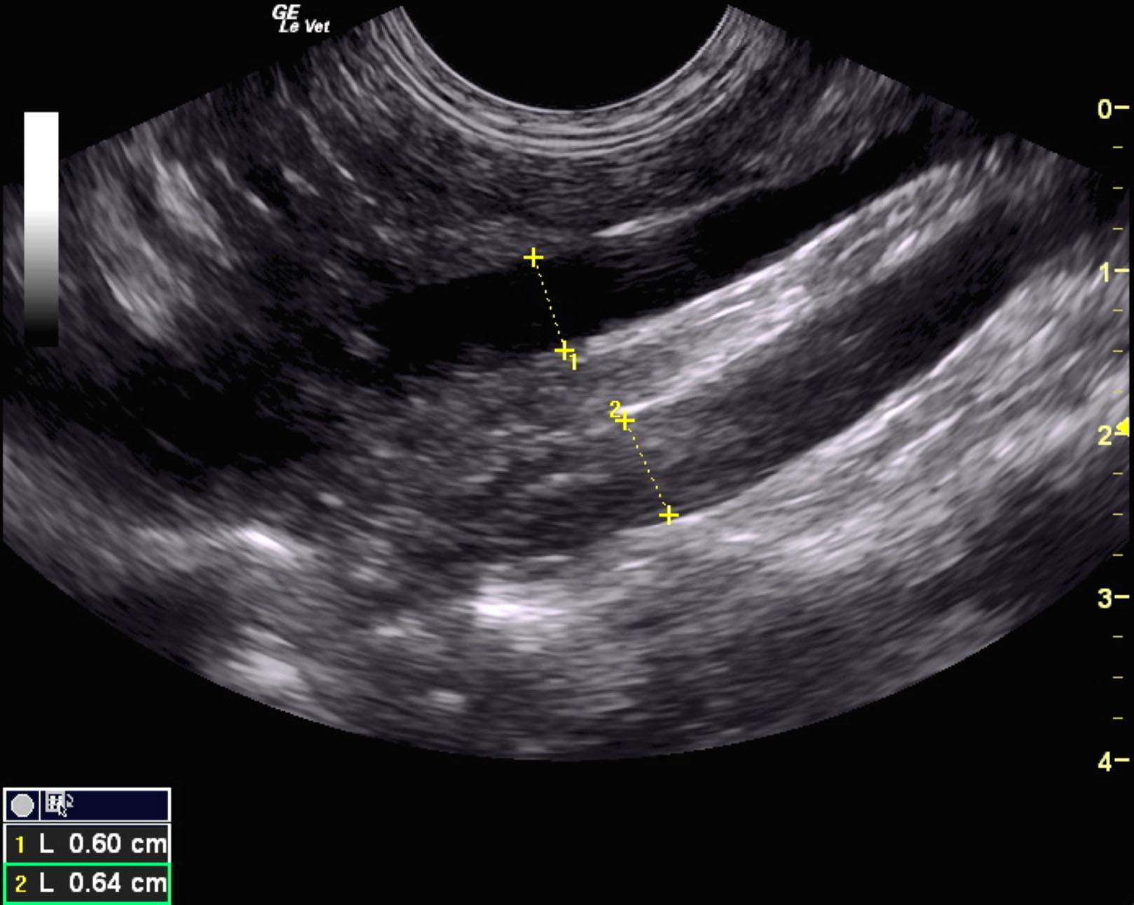
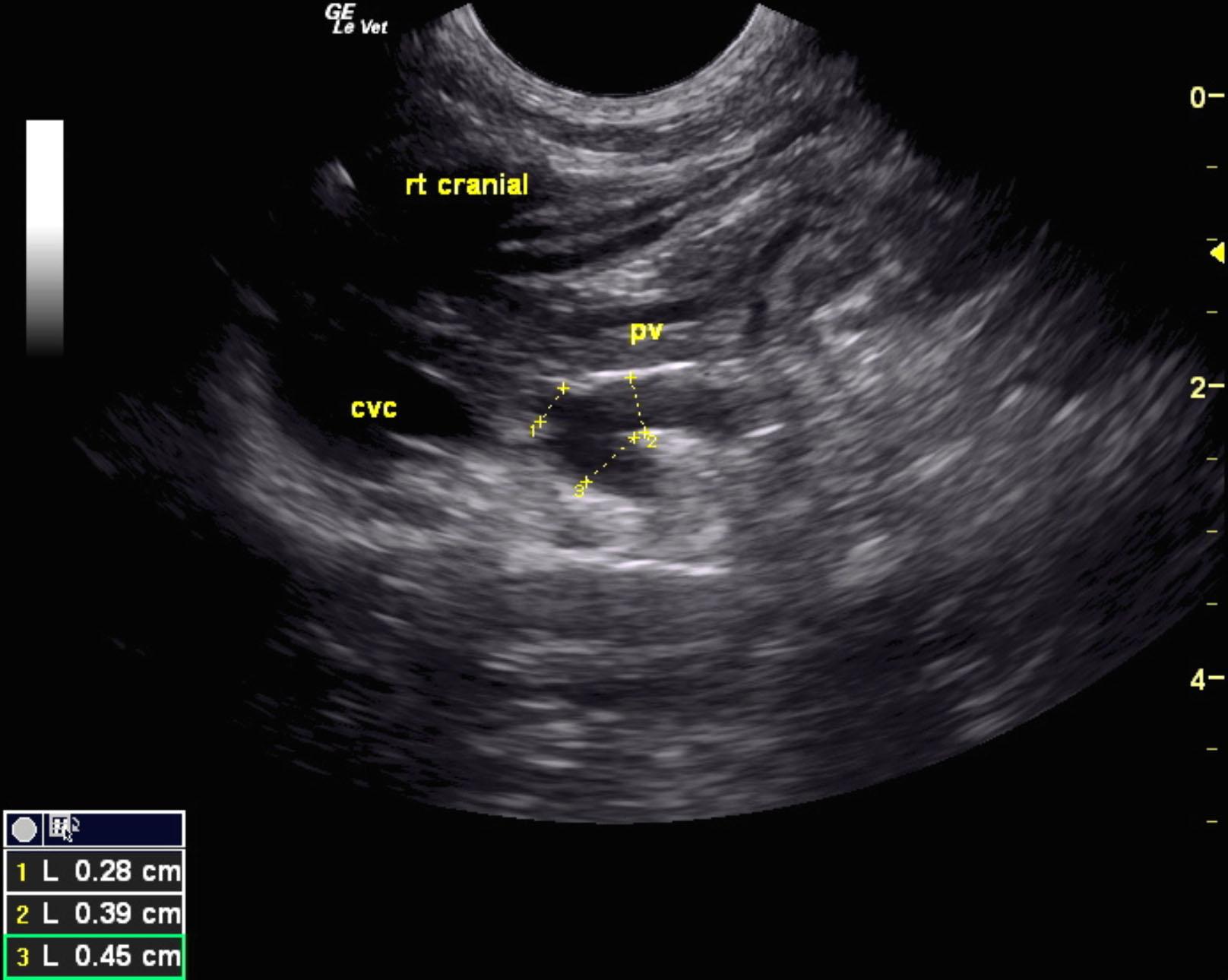
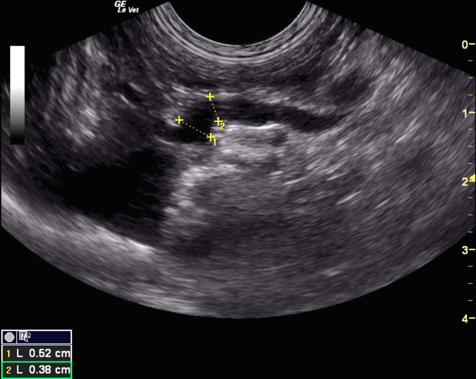
Extrahepatic portosystemic shunt. Splenocaval shunt is suspected.
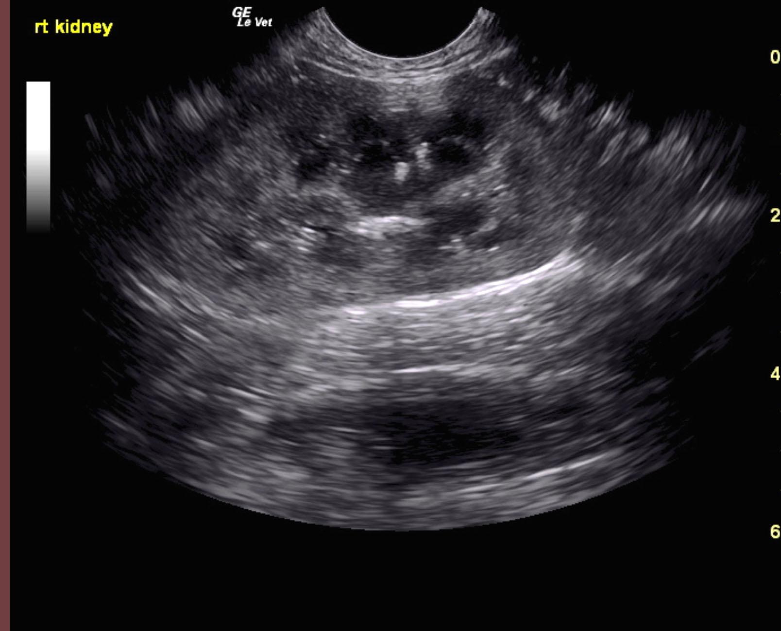
Swollen kidneys with mineralization.
Ameroid constrictor therapy with liver biopsy is recommended. At this time no bladder calculi are present; however, the bladder should be imaged again prior to surgery to assess if cystotomy may be necessary at that time.
Extrahepatic portosystemic shunt was noted in this patient and measured 0.45 cm in width. The portal vein prior to the shunt measured 0.39 cm and post shunt measured 0.28 cm. This is most consistent with splenocaval shunt. The vena cava prior to the shunt entry measured 0.6 cm and the aorta measured 0.64 cm. The extrahepatic shunt passed dorsally and measured from 0.6 to 1.0 cm in width and entered into the vena cava dorsal ventrally prior to the diaphragm. The vena cava measured 1.0 cm at that point. The liver was subnormal in size and mildly coarse in architecture. The gallbladder was unremarkable.
The kidneys were swollen in this patient. The left kidney measured 4.83 cm with medullary rim sign and slight mineralization. The right kidney measured 4.93 cm with dystrophic mineralization and swollen contour.
None
FIP, porto-caval shunt, meningitis
None