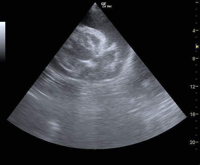
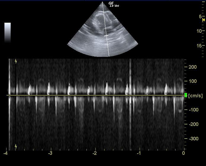
A 10-year-old intact M Vizsla dog was presented for evaluation of one week duration of progressive hacking, non-productive cough. On physical examination increased respiratory sounds, irritable trachea, and mild dental disease was present. CBC was within reference range. On thoracic radiographs marked cardiomegaly with a globoid cardiac silhouette, marked tracheal elevation, and a mild interstitial lung pattern was evident.
A 10-year-old intact M Vizsla dog was presented for evaluation of one week duration of progressive hacking, non-productive cough. On physical examination increased respiratory sounds, irritable trachea, and mild dental disease was present. CBC was within reference range. On thoracic radiographs marked cardiomegaly with a globoid cardiac silhouette, marked tracheal elevation, and a mild interstitial lung pattern was evident.