A 7-year-old spayed female Shar Pei dog was presented for evaluation of seizure activity, secondary to hypoglycemia.
A 7-year-old spayed female Shar Pei dog was presented for evaluation of seizure activity, secondary to hypoglycemia.
A 7-year-old spayed female Shar Pei dog was presented for evaluation of seizure activity, secondary to hypoglycemia.
A 7-year-old spayed female Shar Pei dog was presented for evaluation of seizure activity, secondary to hypoglycemia.
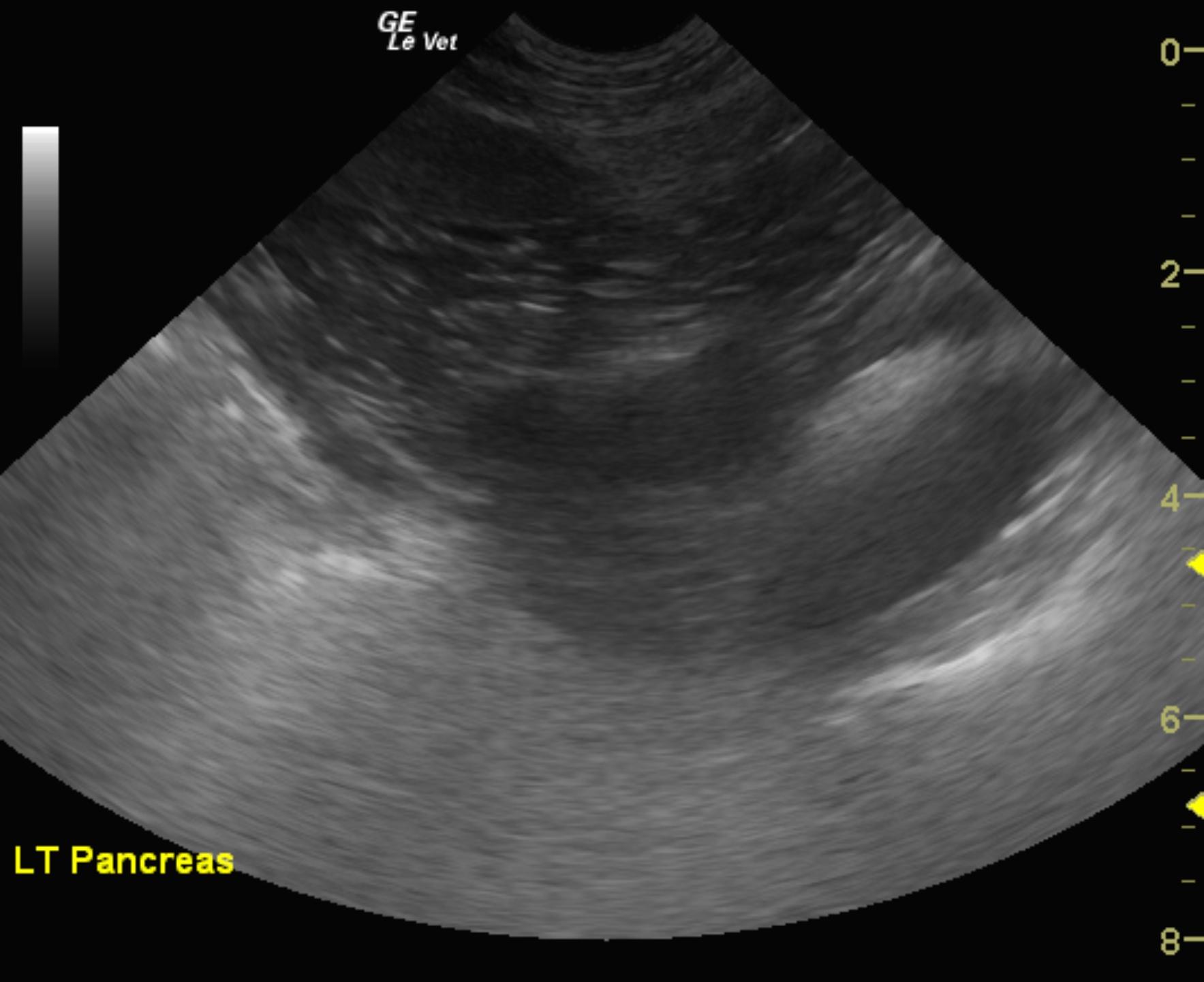
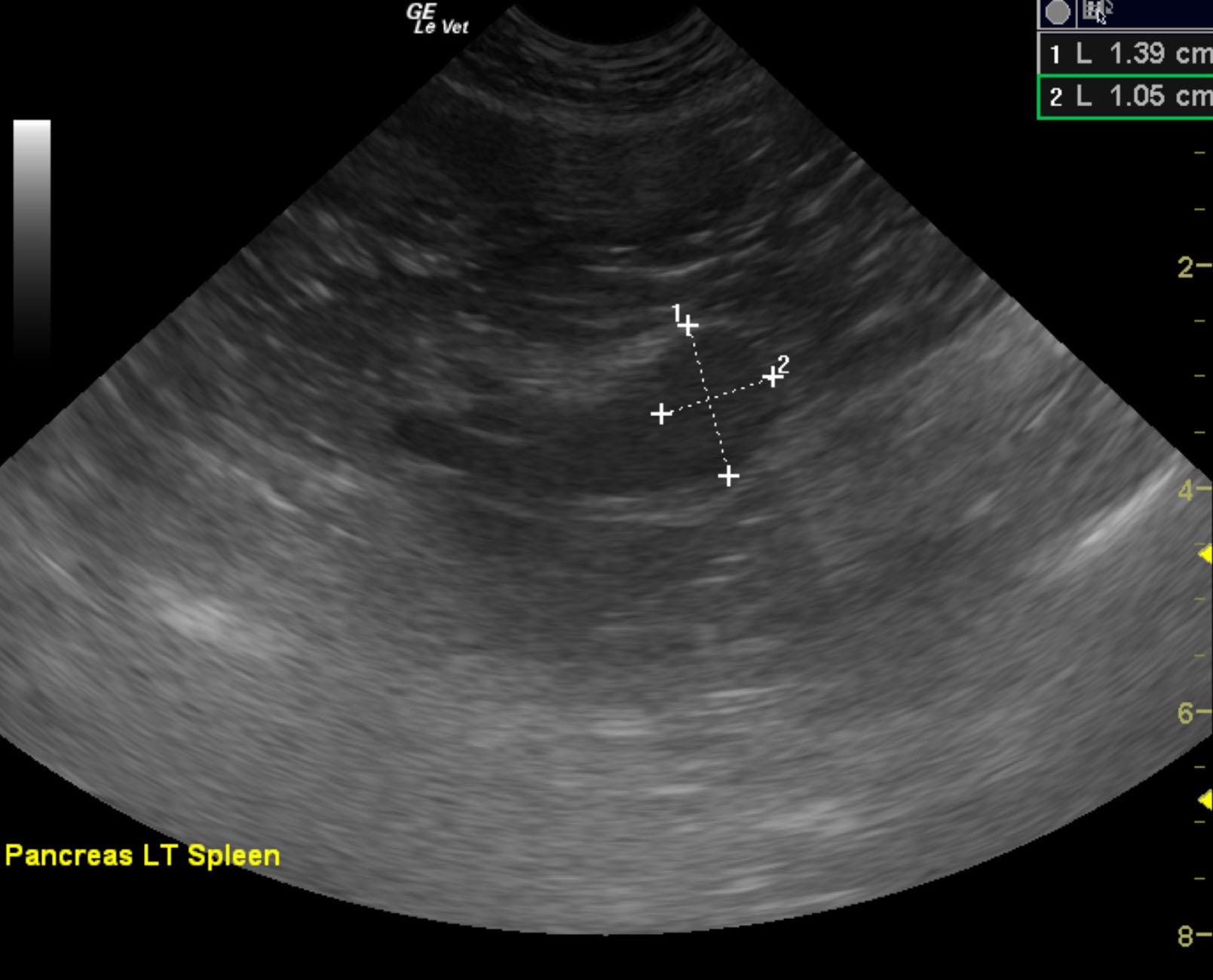
This is consistent with pancreatic nodule at the caudal pole of the left pancreatic limb or potentially a regional lymph node. This is strongly suspicious for metastatic or localized insulinoma. This lesion is adjacent to the mesenteric artery. Therefore, it very well may be a lymph node.
The left pancreatic limb revealed a 2 x 1.5 cm, hypoechoic nodule. The nodule was strongly vascular.
None
Recommend insulin glucose ratio/insulin profile. Surgical intervention and resection is warranted in this patient.
Insulinoma, xylitol toxicity, atypical Addison’s disease, hepatic shunt/cirrhosis, sepsis.
Ultrasound-guided FNAs were performed without complication. FNA revealed a malignant epithelial cell tumor with few clumps of exocrine pancreatic cells.