Prostatic carcinoma in a 14 year old MN Labrador Retriever dog

Case Study

Prostatic carcinoma in a 14 year old MN Labrador Retriever dog

A 14-year-old MN Labrador Retriever was presented for evaluation of pollakiuria and hematuria. The patient had been on Deramaxx for chronic arthritis.

A 14-year-old MN Labrador Retriever was presented for evaluation of pollakiuria and hematuria. The patient had been on Deramaxx for chronic arthritis.
DX

Prostatic carcinoma suspected based on sonographic presentation and clinical history

Sonographic Differential Diagnosis

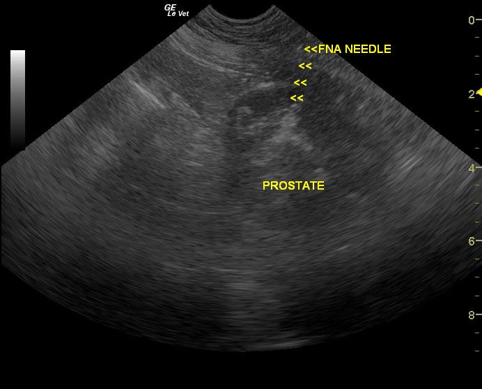
The changes seen to the prostate and enlarged sublumbar lymph node are suspicious for prostatic carcinoma. Fine-needle aspirate or ultrasound-guided biopsy (with coagulation profile) should be performed in order to define the changes from a cellular perspective (via FNA) or structural perspective (ultrasound-guided biopsy).

Image Interpretation

The prostate was visibly enlarged and irregular in contour. Prostatic tissue was patchy with hyperechoic and hypoechoic areas. Areas of mineralization were noted within the prostate. The prostate measured 3.59 x 4.17 cm. Local lymphadenopathy was noted with an enlarged sublumbar lymph node that measured 3.45 x 1.86 cm.

Outcome

The patient was prescribed Deramax and enrofloxacin as a palliative treatment.

The patient was eventually euthanized due to financial concerns and poor prognosis.

Comments


Unfortunately cytology was declined by the owner, so definitive diagnosis of carcinoma was not achieved in this case. However, all indications support a diagnosis of prostatic carcinoma.

Ideally therapeutic options in suspected prostatic carcinomas such as this one, assuming that a definitive dx may be achieved, include repetitive US-guided cyst drainage to minimize recurrent expansive effect of cyst development, treatment for recurrent infection if present by US-guided quinolone injection into abscesses or infected cysts, COX-2 inhibiotor therapy, urethral stent placement, traditional or IR-guided chemo embolization, and radiation therapy.


Clinical Differential Diagnosis

Hematuria – bladder pathology (neoplasia, chronic cystitis, bacterial cystitis, uroliths), urethral pathology (urethritis, stricture, neoplasia, urolith), prostatic pathology (neoplasia, prostatitis).

Sampling

Ultrasound-guided fine-needle aspirations of the prostate were performed. No bleeding was noted post fine-needle aspiration. Urinalysis results revealed urine specific gravity 1.034, pH 7.5, 3+ protein, 3+ blood, WBC >50, RBC >50, transitional epithelia 2-3, squamous epithelia 2-3. Urine cultures revealed no growth. *FNA samples were not submitted by owner choice.*

UA Specific Gravity Range

1.034

Patient Information

Patient Name : Rhett H
Gender : Male, Neutered
Species : Canine
Type of Imaging : Ultrasound
Status : Complete
Liz Wuz Here : Yes
Code : 09_00033

Clinical Signs

  • Hematuria
  • Pollakiuria

History

  • NSAID use

Images

Prostatemeas_11102012122238SublumbarLN_11102012122257ProstateFNA_11102012122319

Clinical Signs

  • Hematuria
  • Pollakiuria
Skip to content