A 1-year-old FS Havanese was presented for evaluation of seizure-like episodes. No heart murmur was present.
A 1-year-old FS Havanese was presented for evaluation of seizure-like episodes. No heart murmur was present.
Case Study
Pulmonary hypertension in a 1 year old FS Havanese dog with seizure-like episodes
Sonographic Differential Diagnosis
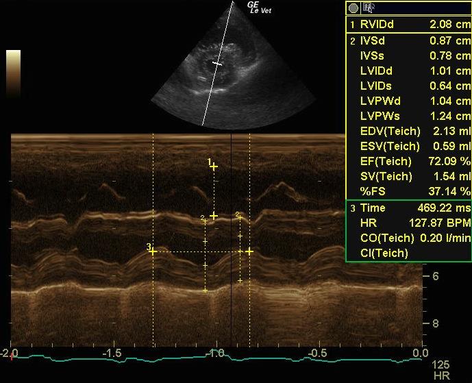
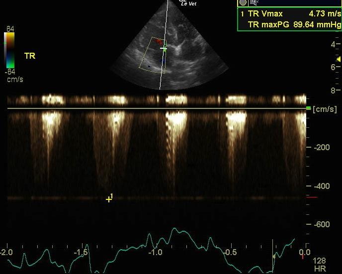
The echocardiogram shows marked right ventricular hypertrophy with a small left ventricular chamber and enlargement of the main pulmonary artery. This is likely due to primary pulmonary hypertension in a dog of this age. The condition appears relatively advanced at this time with the changes noted; this is the likely source of the episodes, particularly if they are seen with exertion.
Image Interpretation
The left ventricular cavity is small in diastole (1.6 cm) and systole (1.0 cm) with a normal fractional shortening (37 %). There is flattening of the interventricular septum in systole, consistent with right ventricular hypertension. The left ventricular walls are relatively hypertrophied (0.65 cm septal wall and 0.65 cm free wall). The right ventricle appears markedly hypertrophied, and the right atrium appears moderately enlarged in size. The main pulmonary artery appears significantly enlarged in comparison to the aorta. There is moderate tricuspid regurgitation on Doppler exam with the jet directed toward the aorta. The velocity is also markedly increased to 4.73 m/sec, describing a pressure gradient of 90 mmHg, consistent with pulmonary hypertension. The heart rate is measured at 124 to 149 BPM and a regular sinus rhythm is seen on an ECG run during the exam.
DX
Pulmonary hypertension
Outcome
Pimobendan at 2.5 mg PO BID and/or sildenafil at 1 to 3 mg/kg PO TID are recommended to try to lower pulmonary artery pressures. No other cardiac medications are recommended based on this exam but exercise restriction is recommended. The echocardiogram should be repeated in 6 months to monitor for any further changes to the cardiac structure. The heart rate should also be monitored to ensure it remains in the normal range.
Comments
Radiographs: The lateral thoracic radiograph shows a mildly enlarged cardiac silhouette with right ventricular hypertrophy. The apex of the heart is lifted off the sternum on this view. The left atrium, pulmonary vasculature and lung parenchyma appear normal. The trachea is elevated over the cranial aspect of the heart.
The patient died after 3 months of therapy.
Clinical Differential Diagnosis
CNS – meningio-encephalitis, congenital defect, hydrocephalus, infectious, metabolic (liver disease, hypoglycemia, electrolyte anomalies), toxins, trauma, true epilepsy. Syncope – cardiac, hypotension, hypertension, hypoglycemia, respiratory disease.
Sampling
None
Video
Patient Information
Patient Name :
Lola P
Gender :
Female, Spayed
Species :
Canine
Type of Imaging : Ultrasound
Status :
Complete
Liz Wuz Here :
Yes
Code :
15_00125
Clinical Signs
- Seizures
Images
Clinical Signs
- Seizures