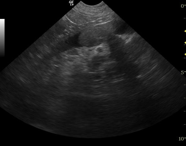
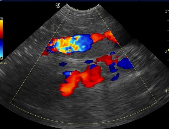
An 11-year-old FS Labrador Retriever dog was presented for the evaluation of polydipsia as well as a five pound weight gain over a five month period of time. The dog was obese and panting excessively on physical examination. Blood chemistry revealed hypoalbuminemia, hyperglobulinemia, hyperphosphatemia, increased ALT, increased AST, hypercholesterolemia and hyperamylasemia. Both lymphopenia and monocytosis were noted on the CBC. The thyroxine serum concentration (T4) was within normal limits.
An 11-year-old FS Labrador Retriever dog was presented for the evaluation of polydipsia as well as a five pound weight gain over a five month period of time. The dog was obese and panting excessively on physical examination. Blood chemistry revealed hypoalbuminemia, hyperglobulinemia, hyperphosphatemia, increased ALT, increased AST, hypercholesterolemia and hyperamylasemia. Both lymphopenia and monocytosis were noted on the CBC. The thyroxine serum concentration (T4) was within normal limits. The urinalysis showed an elevated pH, decreased specific gravity, cloudy appearance, proteinuria, and the presence of bacteria.