Splenic lymphoid hyperplasia, extramedullary hematopoeisis, and a necrotic renal cyst in a 14 year old FS dog

Case Study

Splenic lymphoid hyperplasia, extramedullary hematopoeisis, and a necrotic renal cyst in a 14 year old FS dog

A 14-year-old FS dog presented for a collapsing episode. The owner reported that the patient had been shaking and then fell over. On physical exam, the patient was found to be unsteady on her feet, and she had a rotational nystagmus. Ceruminous discharge was present in the left ear. There was crepitus of a number of joints consistent with degenerative joint disease (DJD), as well as focal alopecia along the dorsal aspect of her neck and tail. The patient was discharged with anti-nausea and ear medications, as well as an NSAID.

A 14-year-old FS dog presented for a collapsing episode. The owner reported that the patient had been shaking and then fell over. On physical exam, the patient was found to be unsteady on her feet, and she had a rotational nystagmus. Ceruminous discharge was present in the left ear. There was crepitus of a number of joints consistent with degenerative joint disease (DJD), as well as focal alopecia along the dorsal aspect of her neck and tail. The patient was discharged with anti-nausea and ear medications, as well as an NSAID. Blood work and a urinalysis were not performed prior to dispensing these medications, however, a urine culture yielded growth of Kluyvera sp. and Escherichia coli.

Blood work was performed several months later and revealed hyperproteinemia, hyperglobulinemia, hypercholesterolemia, hyperamylasemia, as well as elevated SAP and elevated GGT enzyme activities. Thrombocytosis was present on the CBC. The TT4 was within the normal reference range.

Recheck blood work a few months later showed an improvement of the SAP, but the ALT was elevated, and the platelet count was still elevated. The patient did not show any recurrence of the original clinical signs. Approximately one year later, she presented for signs of diarrhea and polyuria/polydipsia. The physical exam showed both nuclear sclerosis and cataracts bilaterally, discharge in the right ear, persistent DJD, as well as muscle atrophy of both hind limbs. The dog was administered subcutaneous fluids, injectable gastroprotectants, antibiotics and anti-diarrhea medications. She was discharged with oral gastroprotectants and anti-diarrheal medications. 
DX

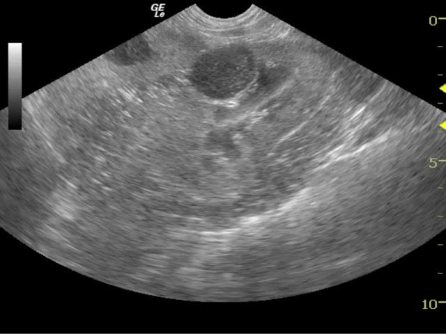
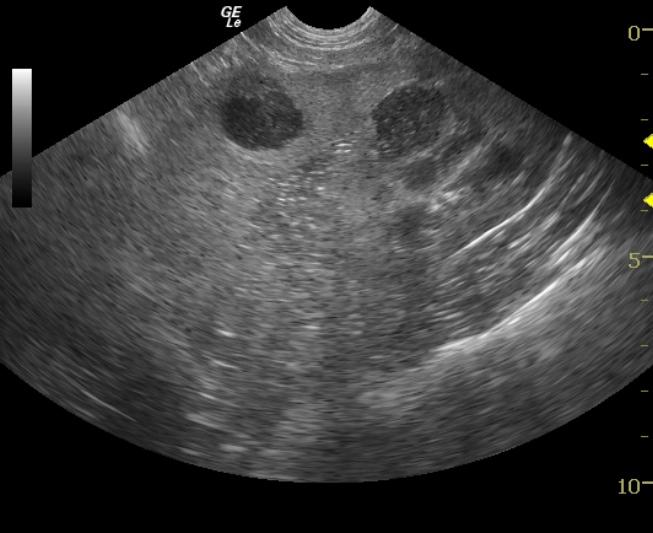
Splenic lymphoid hyperplasia and extramedullary hematopoiesis, necrotic renal cyst

Sonographic Differential Diagnosis

Splenic nodules – abscessations/necrosis, neoplasia (round cell or similar), hyperplasia. Renal lesion – necrotic cyst or abscess, (uniform margins render neoplasia less likely but it is still possible).

Image Interpretation

The spleen presented largely hypoechoic nodular changes with loss of parenchymal detail within the mid body of the spleen. Concurrent hypoechoic necrotic cyst, abscess or mass was present in the dorsal cortex of the left kidney. The lesions are non vascular based on power Doppler assessment. Follow-up of the renal lesion revealed no progression and minimal regression after treatment with antibiotics. However, the splenic lesions resolved completely with only minor echogenic remodeling of the spleen in the region of the prior nodule (11 o`clock position on the 6 weeks follow up video).

Outcome

The patient was stable on follow-up recheck sonograms at 3 and 9 months. The renal lesion remained stable without progression nor regression. Minor splenic remodeling remained.

Clinical Differential Diagnosis

Hyperadrenocorticism, chronic active hepatitis, hepatic abscess, neoplasia, hepatic: hepatoma, adenocarcinoma, lymphoma; gastrointestinal: lymphoma, adenocarcinoma, leiomyoma, leiomyosarcoma, mast cell tumor; reactive hepatopathy. History of UTI, possibly due to underlying hyperadrenocorticism.

Sampling

US-guided FNA of the spleen and kidney. Cytology revealed lymphoid hyperplasia and extramedullary hematopoiesis. The cytology of the renal lesion revealed necrosis. Negative culture on both samples.

Patient Information

Patient Name : Kelly G
Gender : Female, Spayed
Species : Canine
Type of Imaging : Ultrasound
Status : Complete
Liz Wuz Here : Yes
Code : 08_00010

Clinical Signs

  • Diarrhea
  • PU-PD

Exam Finding

  • Muscle Wasting
  • Ocular abnormality

Images

splenic_nodulevlcsnap-2013-07-18-14h47m07s37

Clinical Signs

  • Diarrhea
  • PU-PD
Skip to content