A 3-month-old M American Eskimo Dog was presented at an emergency hospital for seizures. Blood smear evaluation showed mild polychromasia. On CBC moderate anemia and mild thrombocytopenia was present. Serum chemistry showed hyperphosphatemia and hypoglobulinemia. The patient was treated with intravenous fluids spiked with 2.5% dextrose and oral lactulose. The following morning the patient was transferred to rDVM. On physical examination the patient was sedate and quiet, ambulatory on all four legs, with anisocoria.
A 3-month-old M American Eskimo Dog was presented at an emergency hospital for seizures. Blood smear evaluation showed mild polychromasia. On CBC moderate anemia and mild thrombocytopenia was present. Serum chemistry showed hyperphosphatemia and hypoglobulinemia. The patient was treated with intravenous fluids spiked with 2.5% dextrose and oral lactulose. The following morning the patient was transferred to rDVM. On physical examination the patient was sedate and quiet, ambulatory on all four legs, with anisocoria. Whilst hospitalized the patient had a seizure, which was treated with intravenous Valium.
Case Study
Normal liver, portosystemic/intrahepatic shunt rule out in a 3 month old M American Eskimo Spitz dog
Sonographic Differential Diagnosis
No evident portosystemic or intrahepatic shunts. Normal liver. If bile acids are elevated, portal vein hypoplasia, microvascular dysplasia or infectious hepatic disease should be investigated by US-guided biopsy.
Image Interpretation
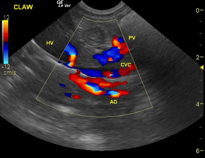
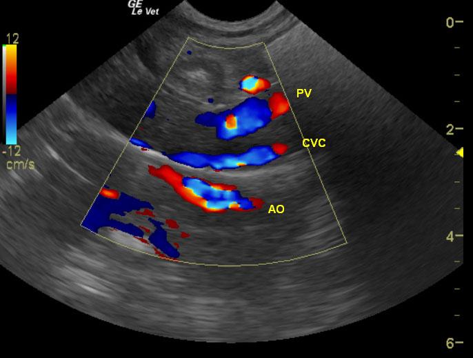
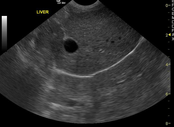
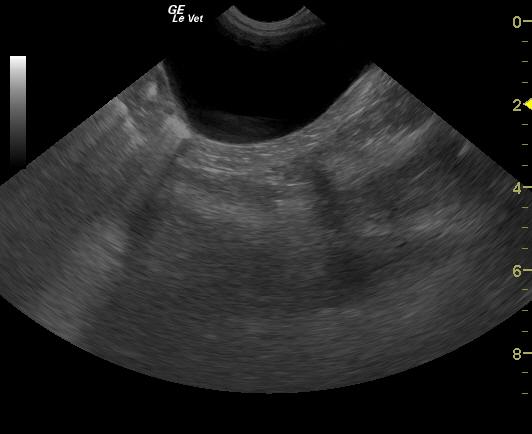
Exam of the cranial abdomen demonstrated normal liver size, contour, and structure. Parenchymal echogenicity was naturally coarse and hypoechoic to the spleen. Vascular and biliary tracts were of normal volume and no evidence of congestion was noted. The portal vein, caudal vena cava, and aorta were 0.6/0.56/0.6cm respectively revealing a PV:CVC and PV:Ao ratio of 1:1 ruling out the presence of extrahepatic shunts. The urinary bladder, trigone and pelvic urethra presented normal wall thicknesses with anechoic urine and normal tone. No uroliths or sediment were visualized.
DX
Outcome
Neurology consult and bile acids panel was recommended based on the sonogram. Preprandial bile acids were within normal range whereas the postprandial sample was elevated. No further outcome or sampling was provided. US-guided liver biopsy would have been ideal to rule out portal vein hypoplasia and microvascular dysplasia.
Clinical Differential Diagnosis
Liver pathology: Hepatic encephalopathy,toxin, acute hepatopathy, hypoglycemia, infectious; meningitis, trauma, congenital defects
Sampling
None taken.
Video
Patient Information
Clinical Signs
- Seizures
Images
Blood Chemistry
- Globulin, Low
- Phosphorus, High
CBC
- Platelet Count, Low
- Polychromasia
- RBC, Low
Clinical Signs
- Seizures