A 3-year-old FS Labrador Retriever dog with history of recent OHE, presented for seizure. Physical exam found patient emaciated, jaundiced, and with decreased skin turgor. In-house chemistry revealed markedly elevated Alkaline Phosphatase, ALT was unable to be determined, hypoamylasemia, low BUN, hyperbilirubinemia, and sample was icteric. CBC found high HCT, high RBC, and low MCHC. ALT analysis was sent out and showed marked elevation. Leptospirosis titer results were negative. Preprandial and postprandial bile acid profile results were both significantly elevated.
A 3-year-old FS Labrador Retriever dog with history of recent OHE, presented for seizure. Physical exam found patient emaciated, jaundiced, and with decreased skin turgor. In-house chemistry revealed markedly elevated Alkaline Phosphatase, ALT was unable to be determined, hypoamylasemia, low BUN, hyperbilirubinemia, and sample was icteric. CBC found high HCT, high RBC, and low MCHC. ALT analysis was sent out and showed marked elevation. Leptospirosis titer results were negative. Preprandial and postprandial bile acid profile results were both significantly elevated. Patient was admitted for I.V. fluid therapy and supportive care. Abdominal radiograph showed decreased detail and small spleen. Coagulation panel run awaiting ultrasound, found prolonged clotting time for PT. Patient continued on fluids, antidiarrheal medication, Lactulose, Vitamin K injections, and antibiotics. Physical exam the following day found patient BAR and extremely icteric.
Case Study
Moderate chronic hepatitis, possible Leptospirosis in a 3 year old FS Labrador Retriever dog
Sonographic Differential Diagnosis
Acute on chronic hepatitis likely. Leptospirosis (endemic region) or similar infection regardless of negative initial titer, toxic insult also possible. Infiltrative neoplasia also possible. Copper storage disease or other forms of chronic active hepatitis suspected. US-guided biopsy is essential for structural diagnosis given the increased portal markings and coarse parenchyma
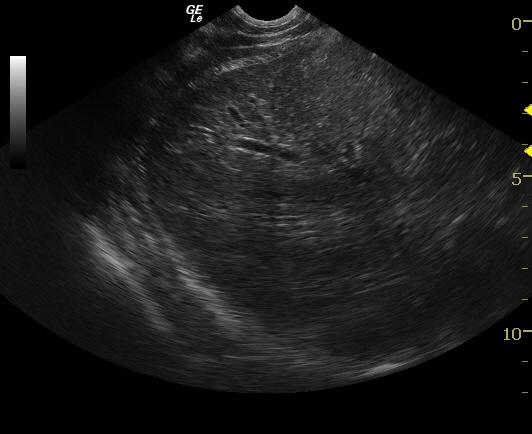
Image Interpretation
Exam of the cranial abdomen revealed normal liver size and contour. The parenchyma did not demonstrate any overt masses. The portal markings were increased with mildly hypoechoic liver parenchyma.
DX
Outcome
Recheck blood work a few days later after IV ampicillin and empirical treatment for leptospirosis revealed hypoalbuminemia, still elevated but improved hyperphosphatemia, much improved ALT, hyperbilirubinemia, low total protein, worsening leukocytosis, monocytosis, neutrophilia, and low MCHC. Antibiotics were later changed and liver protectants were started. Patient began to deteriorate, had pale mucous membranes, was vomiting, anorexic, and lethargic. Later that evening, patient had developed facial edema and fluid rate was lowered. After 1 week of hospitalization, patient was still icteric, on steroids, Ursodial, antibiotics, two gastroprotectants, liver protectant, antidiarrheal medication, and Denamarin, and was still experiencing intermittent, vomiting, and anorexia. Patient was eventually released with oral meds to be given at home, with the plan to check blood values in 1 week. Recheck blood work revealed hypoalbuminemia, hyperbilirubinemia, extreme hyperphosphatemia (4543), extremely high ALT (1938), high AST, high BUN/Creatinine ratio, hyperamylasemia, high GGTP, anemia, leukocytosis, low RBC, neutrophilia, and monocytosis. Over the next several months liver values slowly improved, but patient developed severe edema in left hind leg. Physical exam found severe pitting edema in the left hind leg and distended abdomen; FNA found ascites. Lasix injection was given and due to chronic liver issues, a guarded prognosis was given. Due to the increasing severity of cellulitis in the left hind leg and unresolved icterus, patient was humanely euthanized.
Clinical Differential Diagnosis
Liver pathology: infectious (leptospirosis despite negative initial titer, viral) toxic, inflammatory, immune mediated, hepatic vascular anomaly (intrahepatic shunting congenital or acquired), toxicity (rodenticide, vitamin D), cholangitis, bile duct obstruction.
Sampling
A 16 gauge ultrasound guided biopsy was performed of the liver with no significant complications. Biopsy results taken from liver revealed moderate chronic hepatitis with single cell necrosis, cholestasis and vacuolar change. Sample was also copper stained, but no hepatocellular copper pigment was identified.
Video
Patient Information
Clinical Signs
- Seizures
Exam Finding
- Cachexia
- Dehydration
- Icterus
- Weight loss
Images
Blood Chemistry
- Alkaline Phosphatase (SAP), High
- ALT (SGPT), High
- BUN low
- Post-Prandial Bile Acids, High
- Pre-Prandial Bile Acids, High
- Total Bilirubin, High
CBC
- Hematocrit, High
Clinical Signs
- Seizures
Special Testing
- PT Prolonged