A 14-year-old MN dachshund presented with polyuria/polydipsia. Physical examination was largely normal with excessive body score. Laboratory blood chemistry analysis revealed a moderately elevated SAP and mildly elevated ALT with a normal ACTH stimulation test. Urinalysis showed mild white blood cell (WBC) elevation, bacteria, and a urine specific gravity of 1.018.
A 14-year-old MN dachshund presented with polyuria/polydipsia. Physical examination was largely normal with excessive body score. Laboratory blood chemistry analysis revealed a moderately elevated SAP and mildly elevated ALT with a normal ACTH stimulation test. Urinalysis showed mild white blood cell (WBC) elevation, bacteria, and a urine specific gravity of 1.018.
Case Study
03_00038 Wolfran S Cholangiohepatitis and vacuolar hepatopathy SAVE FOR RESEARCH
Sonographic Differential Diagnosis
Acute inflammatory hepatopathy and neoplasia. Differential diagnoses for the duodenal mucosal thickening include inflammatory disease such as IBD, duodenitis, and less likely, an emerging neoplasia such as lymphosarcoma.
Image Interpretation
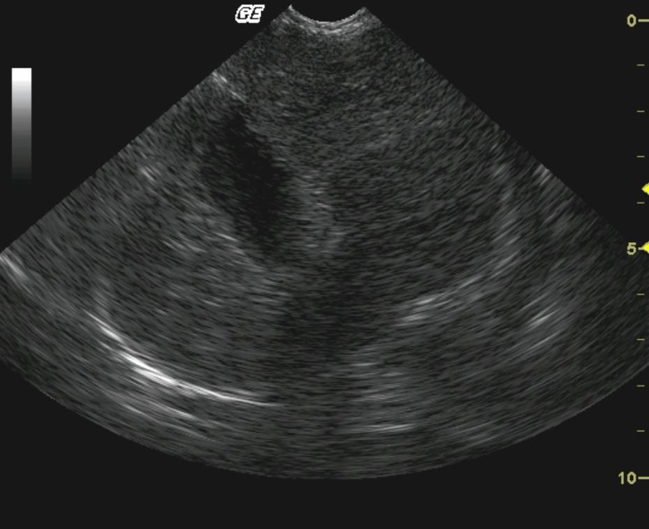
Long-axis view of the right medial liver lobe demonstrates generalized hepatic enlargement. The hepatic parenchyma is diffusely and moderately hypoechoic with a normally smooth and homogeneous echotexture and caudate lobe enlargement. Prominent duodenal mucosa was seen. Video: A cystic lymph node or hepatic cyst was noted adjacent to the portal vein as well as coarse hepatic architecture suggestive for inflammatory hepatopathy. If this is a cystic periportal lymph node then chronic lymph node remodeling and cystic degeneration is likely.
DX
Mild lymphoplasmacytic cholangiohepatitis and vacuolar hepatopathy.
Comments
The purpose of the biopsy was to confirm the suspicion of low-grade hepatic disease possibly related to similar upper GI pathology (IBD) given the combination of prominent duodenal mucosa and low-grade lymphoplasmacytic inflammation in the liver. The definitive cause of the polyuria/polydipsia was unable to be determined owing to a lack of owner follow-up. However, no calculi or masses were evident on the sonogram with moderate degenerative renal disease.
Clinical Differential Diagnosis
Liver: Benign hepatopathy, inflammatory hepatopathy, gallbladder mucocele, neoplasia. Urinary tract: Urinary tract infection (UTI), early renal failure, urolithiasis, neoplasia.
Sampling
US-guided biopsy revealed mild lymphoplasmacytic cholangiohepatitis and vacuolar hepatopathy.
UA Specific Gravity Range
1.018


