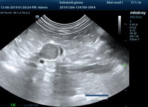
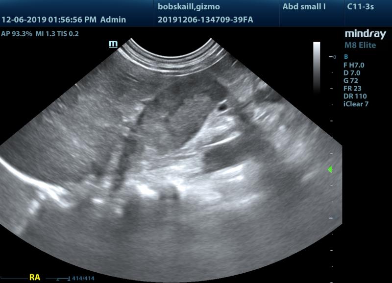
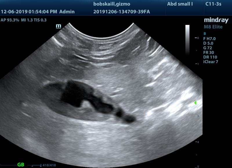
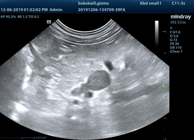
Gizmo is 12 years old, MN, long hair Chihuahua. He presented last week for sudden brusing. Lab showed mild anemia (PCV 35%), Elevated ALP (857) and very low platelets (10K). Abdominal ultrasound showed enlarged adrenal glands (Rt>>>Lt), “something” in the Vena Cava cranial to the adrenal gland and sludge in the gall bladder.
Gizmo was started on Azothioprine for ITP and 1 week later platelets are already >100K. Low dose dexamethasone supression test came back inconclusive (4 hr post – 1.6, 8 hr post 1.1).
Does the “thing” inside the Vena Cava artifact or otherwise? Any thought about the LDDS test result?


Comments
Not a specialist but I’ll
Not a specialist but I’ll take a crack at it. Def not artifact (put color doppler over it and you’ll see blood flow all around it). Those are real nice images BTW!! To me, your ddx are a pheo or ACA in the right adrenal and this mass is invading the phrenic vasculature and vena cava. What you are seeing in the vena cava is either part of the mass or a clot. I think your LDDS is borderline/negative because you don’t have HAC. That’s my 2 cents. Hopefully the experts will weigh in!
Thanks Liz! Note that the
Thanks Liz! Note that the left adrenal gland is quite enlarged as well (especially the caudal pole). I anticipited PTA but LDDS shows differently. Gizmo supposed to be back for BP to rule out Pheo.
Eran
Hey Eran, I saw that, but I’d
Hey Eran, I saw that, but I’d be wondering about either neoplasia in both glands or an adenoma in that left gland. Keep in mind that a BP won’t rule out pheo as the BP can fluctuate dramatically with that particular disease process. Next step would be a CT to eval resectability – some surgeons will cut these but the cases I’ve had with vascular invasion haven’t done well. Edited to add: I have had one of these that was cut by the surgeon only to find out it was too extensive to resect. They biopsied it (pheo), closed the dog up, woke her up, kept her on phenoxybenzamine, and she lived another 2 years with a vena cava that looked like this. So, its certainly not treatment or death, surprisingly.
Interesting Liz! I recall
Interesting Liz! I recall Eric saying they could truck along with something like that in their vena cava, but 2 years is impressive! – Karen
Yeah funny enough the dog was
Yeah funny enough the dog was owned by a neighbor who took her for daily walks by my kitchen window…so I knew she did well with good QOL for those years. It amazed me and became a running joke as I would always express surprise that she was alive probably weekly for 2 years. Haha!
Good to see you guys dont
Good to see you guys dont need me:) nice imaging and cvc invasion of rt adrenal as you can see the mass invasion conncection throught he phrenic in th evideos. Youcan have pheo and pdh together and carcinoma and pdh together…or even bilateral tumors.
The negative LDDS would make
The negative LDDS would make this a non-functional adrenal cortical tumor. As pheo is a possible, can consider looking at urine catecholamines to confirm the diagnosis.
Thank you all for the
Thank you all for the insights! Eran