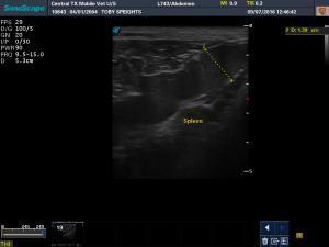
-older DSH, significant vomiting and weight loss over past few months
-no significant abnormalities on CBC, panel, T4
-I found a large spleen and a cystic-type mass with associated enlarged lymph node in the right cranial abdomen. I assumed it was pancreas or less likely liver. The appearance is very cystic but I thought it was separate from the liver.
-FNAs of the mesenteric lymph nodes, spleen, and cystic area were normal. They called the cystic mass normal liver. I really thought I was in it but maybe not? Would a FNA of a cystadenoma come back as normal liver?
-Thoughts on the images? The GI tract was unremarkable. What now? I saved slides for PARR if that seems like a reasonable next step.


Comments
I don’t know if it is on my
I don’t know if it is on my end- but all your images and cines are dark
Can anyone else see the
Can anyone else see the images clearly?
I have had trouble viewing the images from my new machine on my computer but I got a new computer and, at least from my end, I thought that solved the problem. I can view them fairly well on my computer. They are slightly darker than on the machine but not enough to prevent them from being diagnostic. I previously used a Mac and they were always too dark.
Thanks for letting me know but if someone else can let me know how they look on their end, I’d appreciate it.
So I just pulled up this
So I just pulled up this thread on my older Apple computer and the images and cines are MUCH darker than when viewed on my new PC. I’m not sure whether that’s a technical issue on the side of the ultrasound machine or the Sonopath website? I have transferred images to other PC’s and the older ones also have very dark images but they seem to look good on newer ones. Very strange (and frustrating!)…
I uploaded a photo that shows
I uploaded a photo that shows this thread on my Apple vs. my PC computer. It’s not as obvious on the photo, but you can see that the image is much darker when viewed on a Mac as opposed to a PC. Any technical advice? Also advice on the images?
Sonosite machine does this
Sonosite machine does this and has had offloading issues on multiple machines that I have seen. They either come off the machine in huge file ssizes in dicom like 2-3 gb or other times come over dark. We havent had this issue with other people uploading just the sonosite machines. there is something inherent in their software that nobody seems to know what to do wiith. Similar to the logic e sometimes piles on huige file size intermittently for now reason on some software versions when offloading in dicom. Has anyone had similar issues wiht othe rmachines?
re the images I would surely
re the images I would surely fna the spleen at 1.5 cm … any spleen > 1.1 cm in a clinical cat needs a 25 g fna in my book. Looking for round cell neoplasia vs splenitis
Thanks – I did FNA the
Thanks – I did FNA the spleen, it came back as normal spleen. Other suggestions?
The cystic area came back as some slides with normal hepatocytes and some with small well differentiated normal lymphocytes. Would a cystadenoma do that?
Also, just to clarify, my machine is a Sonoscape, which is not the same company as Sonosite (but may have similar issues apparently).
i’m sorry this thread
i’m sorry this thread degenerated into a techinical issues thread but I still would like opinions on the images (sigh, assuming you can see them…)
I have already FNA’d spleen, lymph node, and mass and they all came back normal.
What do you think the cystic area is? Is it likely to be the cause of vomiting and weight loss? Or does chasing lymphoma with PARR make more sense even though the lymph nodes are not really enlarged?
cystic liver area is likely
cystic liver area is likely cystadenoma and fits with the fna results
Cystadenoma unlikley to cause
Cystadenoma unlikley to cause chronic vomiting and weight loss. With normal spleen and lymph node cytology, lymphoma unlikely and thus doing PARR may not give further information. Even though the GI tract looked normal, with the clinical signs, normal blood work, and relatively normal ultrasound, it would an important etiology – scope and biopsies ideal; oherwise hypoallergenic diet, course of fendendazole or metronidazole, and possibly prednisone.
Thanks – I’ll speak to the
Thanks – I’ll speak to the owner and see what he wants to do.