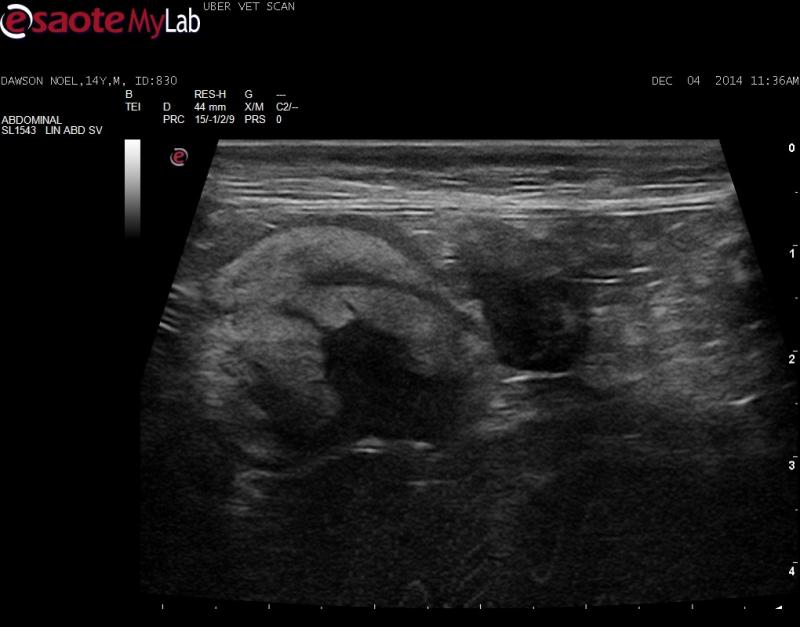
– 14 yr old MN DSH diagnosed with hepatic fibrosarcoma of the left liver lobe about 1 year ago
– liver lobectomy was performed and there was no other evidence of neoplasia at the time of sugery
– pet now losing weight, poor appetite
– there are now masses in the rest of the liver and lymphadenopathy on ultrasound
– in the mid-abdomen there is a hyperechoic structure with an unusual appearance that I am having trouble identifying
Is this neoplastic omentum? Big ugly LN? Something else?
There is an ommental carcinomatosis-like pattern in the rest of abodmen.


Comments
Given the echotexture and
Given the echotexture and position and looks like the mesenteric artery in the middle IM betting on fibrosarc infiltrated mesenteric LN. Likely need to core bx to get the dx because fibrosarc tough to exfoliate on fna… maybe an 18g.
Given the echotexture and
Given the echotexture and position and looks like the mesenteric artery in the middle IM betting on fibrosarc infiltrated mesenteric LN. Likely need to core bx to get the dx because fibrosarc tough to exfoliate on fna… maybe an 18g.
I agree with Eric. Likely
I agree with Eric. Likely mets to the mesenteric lymph nodes. Will take agressive needling while applying negative pressure to the syringe while stabbing to get cells. Usually I get results on these kinds of lesions with this technique.
I agree with Eric. Likely
I agree with Eric. Likely mets to the mesenteric lymph nodes. Will take agressive needling while applying negative pressure to the syringe while stabbing to get cells. Usually I get results on these kinds of lesions with this technique.
Nice technique marty! I go
Nice technique marty! I go bigger needle and corkscrew the needle using th ebevel as a cutting device carving into the tissue when its really dense.
Nice technique marty! I go
Nice technique marty! I go bigger needle and corkscrew the needle using th ebevel as a cutting device carving into the tissue when its really dense.
Interestingly in this case I
Interestingly in this case I did both an FNA and 18 gauge core biopsy on the original lesion in the liver with both coming back inconclusive. The lesion was extremely firm/dense. It was not until we removed the liver and sent a large chunk of tissue for histopath before we could get a definitive diagnosis.
So I agree, fibrosarc is hard to biopsy. Likely will not go further with this case as the lesions are so wide-spread, and knowing that the patient had fibrosarc aready previosuly confirmed.