Dear All,
I need Your help again in this “small liver” patient. She is 7 years old. She had epileptic seizures- elevated liver enzymes.
No sign of urinary stones. Liver function tests ( BA, ammoniak) pending.
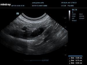
US shows smal liver size, distended gallbladder, small v portae/ v cava ratio ( 0,75/1)
In transversal view i can see a blue vessel which flows directly to the v. cava. I could not image it in sagital view.
I think this could be a splenocaval one- but i am not sure.
Thank You!
Rita

Comments
Certainly a small liver
Certainly a small liver worthy of an ehpss but Im not sure what is going on here on the vasculature.What you always want to shoot for is the obtain the cvc/ao ratio that would be right intercostal for sure on this dog because of the severe microhepatica (posiitons 11-13 sdep abdomen https://sonopath.com/products/downloadable). Then if its a shunt entering the cvc then the cvc will be big and turbulent (on CF) and from there take videos around the turbulence working toward the pv area.
Check out this youtube link I did a while back
https://www.youtube.com/watch?v=YgUPzGB-jpI
Heres a shunt search from the sonopath archive
http://sonopath.com/members/case-studies/search?text=shunt&species=All
Here’s a shunt search in the sonopath forum
https://sonopath.com/forum?keys=shunt
and here’s our shunt research lecture for ecvim 2010
http://sonopath.com/resources/research-publications
When in doubt send to CT as you can see some in our sonopath archive diagnosed on CT which is the most sensitive method but of course more expensive:)
Attached is the normal portal hilus for reference.
I have read these
I have read these articles-are all very useful!
I took pictures from the hilus, measured the 3 vessel-but could not upload the image.
I will try-just i am not at home now.
V portae was 70% of the v. cava.
I will opload the other images!
Rita
Dear EL,
I have found tgis
Dear EL,
I have found tgis measurement-but no video from the v cava.
I will take it next time, when he comes back
Thnak You!
Rita
You can always do the
You can always do the micro-bubbles study for PSS (read this https://www.vetoclock.com/wp-content/uploads/Microbubbles-Portosystemic-Shunts-VET-RAD-ULTRA-CV.pdf)
If there is a macroscopic shunt you should see the micro-bubbles in:
– Pre-hepatic CVC + Abdominal Post hepatic CVC + Right Atrium: ehpss
– Abdominal Post-hepatic CVC + Right Atrium: ihpss
– Only Right Atrium: porto-azygos shunt
Very easy and safe to do, but I’m with EL, you should always confirm with CT for surgery purposes.