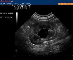
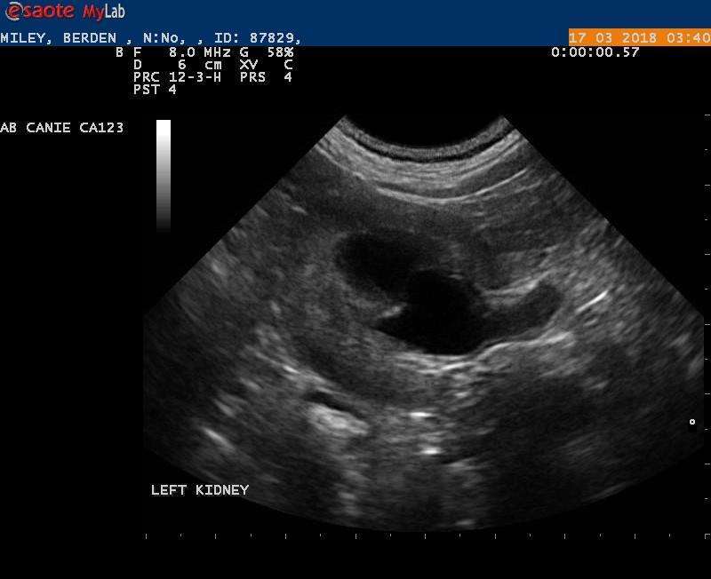
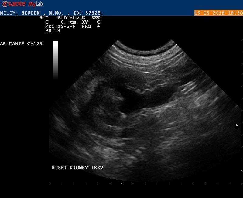
Millie is a 2 years old Pug that initially presented for straining to urinate, blocked on October 2017. She was diagnosed with bladder stones and had a cystotomy done .According to owner she is incontinent ever since the surgery; she is still urinating with a good stream but also dribbling urine ( wearing a diaper..)
Her bloodwork ( CBC, Chem, lytes) is normal but BUN, Creat is high normal
U/S showed normal bladder wall ( no suture reaction, no TCC), intravesical ureters seen on doppler after Furosemide inj.Bilateral hydronephrosis evident but no definitive urolith seen while scanning the dilated ureters or bladder.
I wonder if this presentation can be pyelonephritis as was suggested by a radiologist ( teleradiology); because I never seen one so bad… or this is most likelly due to stricture, other. If partial obstructed by ureterolithiasis would I still see good ureter jets?
Thank you,
Calin
Forget to add that she had UAs, C/S that was negative, and she was on antibiotics 2 tx courses and currently on PPA that doesn’t help


Comments
anybody ?
anybody ?
Hmm I replied to this on the
Hmm I replied to this on the 17th but it didnt stick for some reason..
On the vidoe it looks like there may be an ectopic ureter or ureteral dilation from stricture. Would need to follow it into sdep position 3 deep urethra to see where the straining and incontinence issues are as these are usually lower urinary issues. The kidney looks like hydroureter possibly from a stricture or congential malformation. Pyelonephritis may be there but no inflammatory patterns here just pelvic dilation which is not synonymous with pyelonephritis. This is more of a structural issue to me based on your image sets and not likley a primary inflammation issue such as pyelonephritis. Need to dig into the diagnostics further such at more us images as described above or CT with contrast or even IVP with rad series.
Do a vaginal scope as well to look for distal abnormailites.
I ve seen both ureteral jets
I ve seen both ureteral jets in the bladder, one maybe more apical then I would expected but definatelly not in the urethra. Pelvic urethra was normal based on US. we ve recommended a CT, not sure if owner would follow through. I was just curious if can call it pyelo based on kidney appearance. I heard some people talking about the symetry of pelvic dilation to differentiate fluids versus pathologic?
No you can’t call pyelo with
No you can’t call pyelo with pelvic dilation only. Technically the only way to truly call it is with pyelocentesis and urinarlysis from the pelvis. With dilated ureter like yours then its hydronephrosis-minor because of distal obstruction… lilely strictured ureter in this case for whatever reason. If the pelvic fat is ill defined suggesting inflammation and the pelvic urine is echogenic then this would fit pyelonephritis and you can match it with pyuria (urinary wbc > 5 with concentrated urine or less with dilute urine) in the urine from the cysto or from the pyelocentesis. But pyelectasia/hydro can be caused by a number of things and especially scarring from insults/stone passage, surgical strictures and idiopathic. Pyelectasia from fluids is theoretically bilateral, fits with history of fluid tx while doing the sonogram and not more than 3-4 mm dilation.
make sense. Thank you EL.
make sense. Thank you EL.