- 10 year old mn DSH presented for chronic occult and frank hematuria
- Urinalysis showed hematuria w/o bacteria or crystals. SpG 1.050, pH 7.0
- Nonsresponsive to antibiotics
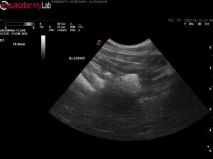
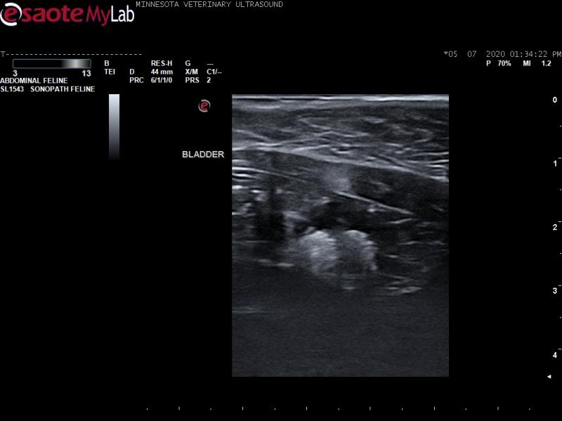
- Abdominal ultrasound showed strong shadowing echogenic densities in the bladder lumen suggestive of bladder stones.
- Surgical cystotomy revealed a lobulated bladder mucosal wall mass and diffusely thickened bladder wall. The primary vet said that the tissue was very tough to cut through.
- Histopathology performed on the bladder mass biopsies showed ulcerative, lymphocytic chronic cystitis with proliferative fibrovascular tissue, edema, segmental mucosal hyperplasia with Brunn’s nest formation.
- Just wondering if these images look like stones or a mass to you and if any of you have seen this before. The primary vet was unable to resect the whole mass.
- I am also wondering why it looks so hyperechoic….could this really just be due to fibrous tissue or could stones have been missed at surgery?


Comments
Sand, small stones, sand, and
Sand, small stones, sand, and mucous together. Be sure to scan again right before cystotomy as things change in these scenarios of the feline UB.