8 yo MN Pitbull seen at the ER last week for dyspnea/coughing. Severe pleural effusion. 1.6L removed, transudate.
Normal bloodwork.
Pitting edema all 4 legs.
Today, P is doing better, still some pleural effusion, no pericardial effusion, normal heart on brief echo.
Scant peritoneal effusion.
Right pancreas was thickened (2cm), irregular, with a dilated vessels. FNA pending (25G).
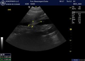
1cm echoic structure in the CVC near the R adrenal: clot or mass?
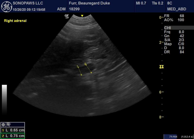
Adrenals looked WNL to me, but I know it could still be invasion form R adrenal…
Thank you!
Julie


Comments
With the pleural transudate
With the pleural transudate and pitting edema would worry about hypoalbuminemia, which would result in loss of AT III with potential thrombosis formation. Is the reported blood work correct?
Hello Dr. Lobetti,
His
Hello Dr. Lobetti,
His albumin was normal (3.4), which is so surprising with that amount of edema!
Julie
Would recommed that it be
Would recommed that it be looked at again. Other possibilty would be vasculitis.
Will pass the info along to
Will pass the info along to the rDVM. Thank you!
Images are a bit dark on my
Images are a bit dark on my end but i think that us an oblique view of the cvc and overlying mesentery and not a thrombus. With normal albumin and that hostory I’d be worried about lymphoma or lymphangiosarcoma. CT of the pelvis/limbs and chest after draining the chest would be the next step. Cytospin the last part tapped off the chest (more cellular part of the pleural effusion) and slide our the sediment after spin down and look for lymphoma cells or similar but if lymphangiosarcoma may not exfoiate well. The Thoracic duct may be plugged as well as pelvic lymphatics. Could also full thickeness biopsy the edematous part of the legs sometimes it takes that to dx lymphangiosarcoma. Its a zebra dx but its out there and these clinical signs fit.
Thank you Eric. The ER did
Thank you Eric. The ER did the cytology, but I don’t know what technique they used. I will keep that in mind for the next effusion. Do you stain it like a urine sediment?
I passed the recommendations to the rDVM. He had no swollen LN in the abdomen, submd/axillary/inguinal popliteal. Owner can’t afford more tests, so he is on pred/lasix.
Just diff quick or leave
Just diff quick or leave unstained for the lab to stain it.
Our telecytology protocol may help here:
We use this exclusively in our mobile operations.
Thanks for the info!
FNA of
Thanks for the info!
FNA of the pancreas was “low grade mixed inflammation”.
Great on sticking the
Great on sticking the pancreas. It allows for more targeted theray when coming up non neoplastic inflammatory changes as you know if its LP inflammation as opposed to neutraphilic then thats when pred/hydrolyzed diet can be effective in pancreatitis tx. We dont stick the pancreas enough for some reason but loads of info on a nebulous disease especially in cats. With solid technique I and my colleagues have never had a complication sticking the pancreas.
Remo sent me these a while back in his reseach so I will pass it on:
Sampling the pancreas
Crain SK, Sharkey LC, Cordner AP, Knudson C, Armstrong PJ. Safety of ultrasound-guided fine-needle aspiration of the feline pancreas: a case-control study. J Feline Med Surg. 2015 17(10):858-63.
The safety of fine-needle aspiration (FNA) of the feline pancreas has not been reported. The incidence of complications following ultrasound-guided pancreatic FNA in 73 cats (pancreatic aspirate [PA] cats) with clinical and ultrasonographic evidence of pancreatic disease was compared with complications in two groups of matched control cats also diagnosed with pancreatic disease that either had abdominal organs other than the pancreas aspirated (control FNA, n = 63) or no aspirates performed (control no FNA, n = 61). The complication rate within 48 h of the ultrasound and/or aspirate procedure did not differ among the PA cats (11%), control FNA (14%) or control no FNA (8%) cats. There was no difference in rate of survival to discharge (82%, 84% and 83%, respectively) or length of hospital stay among groups. The cytologic recovery rate for the pancreatic samples was 67%. Correlation with histopathology, available in seven cases, was 86%. Pancreatic FNA in cats is a safe procedure requiring further investigation to establish diagnostic value.
There is also tons of information in the human literature – one such example:
Forty fine needle aspiration (FNA) biopsies of the pancreas were performed on 37 patients with a radiologic suspicion of malignancy; 32 aspirations were guided by ultrasound, 2 were guided by CT, and 6 were obtained intraoperatively. A pathologist read a rapid-stained smear of the initial aspirate as the procedure was performed and triaged specimens for routine cytologic, cell block and ultrastructural study in solid lesions plus carcinoembryonic antigen (CEA) assay and amylase study in cystic lesions. Purulent material was studied by gram staining and culture. The overall sensitivity in the series was 81%, with a specificity of 100%. No complications were noted. Ultrastructural examination improved the diagnostic accuracy in two cases. Assays for CEA and amylase in “cyst fluids” differentiated true cysts and cystadenocarcinoma from pseudocysts. Maximum utilization of the material aspirated was useful in diagnosing the etiology of solid and cystic pancreatic masses.
Thanks for the papers!
I used
Thanks for the papers!
I used to never poke the pancreas. I listened to several of your CE lectures a few months ago, and I learned so much. I also use the 25G 1.5 inch needles you recommended. Thank you for that too!