- 12 year old Lab with sudden onset of abdominal distension that comes and goes as per the owner
- Patient has a long history of ingesting multiple FB’s
- The liver is hyperechoic with nodular hyperplasia
- There is evidence of some copper storage disease on previous hepatic biopsies
- Cushing’s disease is suspected ( especially with the polyphagia) but the owner will not allow for a proper work up.
- Ultrasound findings that are of particular interest are the following;
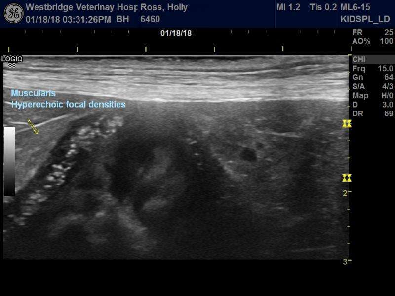
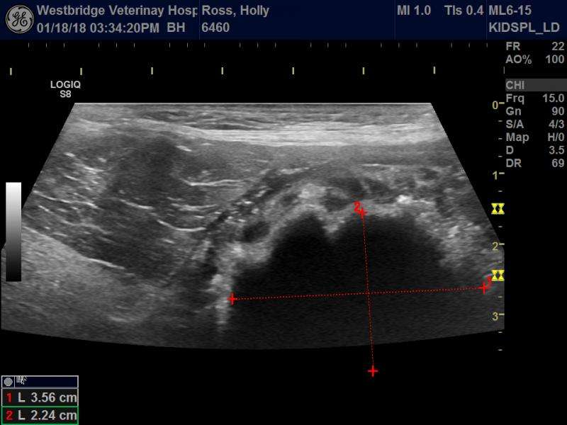
- evidence of a large foreing body in the stomach
- Hyperechoic focal densities present in large accumulations within the muscularis layering of the stomach
- the question is the following; have others seen this in a similar case. I believe that this may be due to the possibility that this is a secondary to the long term presence of the FB. We do not know how long the FB has been there. My suspicion is that it has been there for under two weeks time. This matches the clinical observation timeline wise. The gastric mucosa is very thickened and inflammed. The gastric motility is also very hypermotile. It seems strange to me that we can get such dramatic changes to the muscularis if it has only been 2 weeks since injesting the FB. Could this be totally unrelated? Could this be secondary to the hapatic abnormalities? Could this be older lesions associated with a previous FB?
- unfortunately I forgot to do a Twinkel test and elastography to see if this is mineralization or fibrotic changes.
- I will get an opportunity in the future to re-scan the pet and I will attempt to do these extra images at that time.


Comments
Wonderful image set as always
Wonderful image set as always Bob. The pattern I think is less consistent with fibrosis given the irregular up and down and spotty pattern like mucosal speckling but this is in the muscularis. Potentially more linked to dystrophic mineralization from possible cushings like we see in spleens and renal cortices but I havent had anyone give a definitive on this. Would be great for intraoperative US and get histopath and publish it.
Nele do you have anything recent in your radiology world on this phenomenon? Maybe somethign out of EVDI?
When I see this dog again I
When I see this dog again I will try imaging it with the twinkle setting that seems to help differentiate between mineralization and more organic tissue. I use it in bladders a lot. The other is elastography. It may indicate how firm it is. I was thinking along the Cushing’s line as well but physically this dog does not present as such. She is a very thin dog. The polyphagia and hyperechoic liver keeps on the list of R/O. I will see if we can rule it out with a urine cortisol reading. The owner is not willing to commit:(
The attached image is an example of the Twinkle setting. I will try and keep you updated.
What a perfect example of
What a perfect example of what we talked about in Toronto in our MSK course lab, Bob. I still believe this is fibrosis. I`ve only seen this in cases with a longer clinical history. Overall quite heavy gastritis in this dog for a FB “only”. Do you agree there are focal hyperechoic echoes in the submucosa and mucosa too? even if less than the striking changes in the muscularis. Possibly also some hypertrophic pyloric changes.
I only know of one report of hyperechoic lesions in various layers including the muscularis in the human and canine stomach shown by endoscopic US and proven to be fibrosis by means of histology. But its an old paper and the lesions were placed intentionally to treat LES reflux. SO Im not sure of the value in this context. At elast it shows that muscularis fibrosis in the stomach can cause dense echoes.
Nele
The course was a lot
Nele
The course was a lot of fun and you were wonderful as a teacher. I really enjoyed the conversations. I purposely placed a video of the pylorus to demonstrate that at least to me, that there was no significant hypertrophy at the level of the valvular junction. I see what you mean by some focal densities being imaged in the sub mucosa. I will review some of the other cineloops to see if we captured it better. I also lean more towards fibrosis over mineralization.This FB may have been there for months. We just don’t know for sure. He did not regurgitate it when we administered apommorphine. I will keep you updated.
Thanks Bob, I really enjoyed
Thanks Bob, I really enjoyed it too!!! Likewise & I hope we can repeat it soon
Any possiblity that there is
Any possiblity that there is renal disease in this dog? as I have seen this with uremic gastritis.
They kidneys were both normal
They kidneys were both normal as where their biochemistry values.
I thought that I would attach
I thought that I would attach another video for Nele to show that the changes are limited to the muscularis layering alone. Sorry the program wouldn’t let me.
The image is a screen capture outlining the restricted area where the fibrosis is found. The drawing tries to demonstrate the directional path that the FB would be subjected to with the force of the gastric contractions. Because I suspect that this object has been in the stomach for a long time it has had time to alter that portion of the pyloris. The changes in the muscularis may illustrate its attempts unsuccessfully to eject the object out from the stomach.
You`re an artist Bob! Makes
You`re an artist Bob! Makes sense to me. BTW I just had 2 super cool MSK cases where we have correlative images of MRI and US plus biopsies: one myositis of the shouolder girdle and one presumed (biopsy result pending) sciatic nerve myositis where we took surgical biopsies of the nerve 🙂
Will you share those images
Will you share those images of the sciatic nerve and myositis? Super cool cases from you mean really amazing, now you have me curious. Today, I’ve only had an US diagnosis of pneumonia, another classic triaditis, one with severe IBD with major lymphoid hyperplasia within the sub mucosa of the ascending colon and ielum and finally a recheck of a strange lesion in the jejunum of a cat who was treted for intestinal lymphoma over 5 years ago. Hey the day is still young.
I’m worried that I will quite effectively drive both yourself and Eric nuts over time as we all live and breathe US. It is actually such a pleasure to have these kind of discussions with people such as yourself and Eric. You both have such a passion for it and all of us just want to advance the art even further for the benifit of all.
I feel like Im a spectator at
I feel like Im a spectator at the sonographic nerd olympics and totally enjoying it:) Great to have you in the forum Bob… and Nele of course your brilliance is always appreciated!
Eric
Is there a medal
Eric
Is there a medal involved? It can’t be gold. It would have to be grey as we are often referred to as the shadow people. We are all just playing for the love of this sport! It is fun to be here :). Looking forward to other discussions. You have a great team,Bob.
Bob I just posted the sciatic
Bob I just posted the sciatic case in the forum. Not sure how to alert you. Eric can you help Bob find it? Video was too big file size unfortunately because its really cool…:/
Its comes up listed on the
Its comes up listed on the left or use the search for it on the left column or Bob here is the link:
https://sonopath.com/forum/msk-us-sciatic-nerve-neuritis
nele if you send me the file wetransfer I can convert it or use handbrake to convert monster files to mp4 or i like Movavi converter at 69$
its mp4 but its huge more
its mp4 but its huge more than 500MB. You wanna try?
Nele, thank you for the heads
Nele, thank you for the heads up. I will ask some questions on your blog. I’m going to post another interesting case from yesterday. Would love some feedback. Have a wonderful day.