hi everyone,
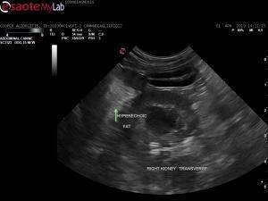
Just wanted to know if anyone has had similar cases to the images I had and what your thoughts are. I scanned this dog and subjectively felt the peritoneal fat surrouding the major organs were hyperehoic. (would you agree or am i overseeing things?) There is NO free abdominal fluid.
hi everyone,
Just wanted to know if anyone has had similar cases to the images I had and what your thoughts are. I scanned this dog and subjectively felt the peritoneal fat surrouding the major organs were hyperehoic. (would you agree or am i overseeing things?) There is NO free abdominal fluid.
This is a rather strange case; 7y.o MN beagleX cavalier that presented with acute lethargy. He had pericardial and pleural effusion which resolved after a couple of days ( no masses and ECHO was normal). There were bruising noted subcutaneously as well from blood taking. APTT and PT were within normal limtis but buccal mucosal bleeding test was prolonged. Can this be some weird vWF disease? have you seen dogs with primary haemostasis disease with hyperechoic fat? how about immune mediated disease? vasculitis?


Comments
Bit old for vWF disease to
Bit old for vWF disease to set in. Besides vasculitis, shoud also consider steatitis.
Thank Remo. How about
Thank Remo. How about microscopic bleed within the mesentery? Would that look like this? I’m trying to make sense of his clinical signs and sonographic findings. Would steatitis occur in a patient with primary haemostatic disease?
Vaculitis would be a cause
Vaculitis would be a cause for mesenteric bleeding. Steatitis not associated with primary hemostatic disease.