– 10 year old DSH with a loud murmur and syncopal episodes
– HCM with dilated left atrium and an arrhythmia
– I have a few questions please?
– I think I am seeing VPCs on ECG. Holter is not an option, do you think ventricular tachycardia is cause of collapse? Would you trial sotalol?
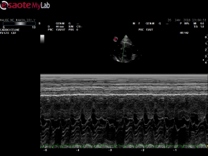
– On m-mode at level of mitral valve, is this a separated e and a wave rather than SAM? I want to rule out LVOTO as cause of collapse
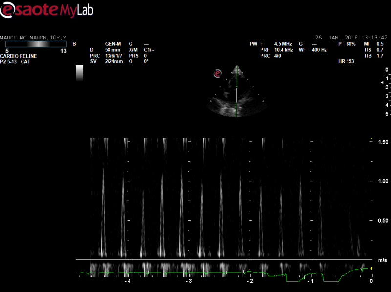
– On mitral inflow there is an increased velocity of summated e and a waves, can you say anything about diastolic function or just increased left atrial pressure?
– I have 2 vidoes of colour doppler on AV and MV with lower and higher scale. Is this just aliasing at lower scale? I often lose colour at higher scale and find it difficult to locate source of murmur. I try to decrease frequency, window of b mode and colour but often no change, does anyone else have this problem with 5-13 probe on my lab alpha?


Comments
Ok- I will commit my
Ok- I will commit my opinion.
Hypertrophic cardiomyopathy, grossily dilated L Atrium
A wave larger than E wave indicating diastolic dysfunction.
SAM of the anterior mitral valve leaflet.
I would not buy any “green bananas” for this cat.
Screen shot of the last frame of the 3rd video- indicating SAM
OK- lets see what others have to say.
I think yiou have a
I think yiou have a combination of issues here. Arrythmias need to be > 20-30/minute or in runs to cause clinical signs as a rule. Even though frequent in these clips if the arrtyhmia is assumed similar thoughout the day then the arryhtmias is a side effect of myocardial strecth/hypoxia/irritation. But this is a big assumption since paroxysmal events can certainly occur. From apical view looks like both fixed lvot obstruction and dynamic from 5 chamber view. Volume overload as well. I always grew up in the cardiology school of tx the volume overload first and see what th earryhtmia does. Also never slow a cat heart with obstruction and volume overloiad since they need the elevated HR to maintain themselves… again tidbits from my cardio hx.
I would tread lightly here and use lasix and plavix only for 48 hours with cage rest and reecho/ecg to see how the heart is responding. Then once the volume is more under control we see about rate tx. Im sure 10 people may do 10 different things here but this is what I would do. Lets see what CardooMcGyver Peter would do here:)
FYI Peter Modler (https://sonopath.com/about/specialists/peter-modler-dvm-dipl-tzt) is lecturing at our October full sdep echo lab or if you want a warmup SDEP technique for normal aquired and congenital seminar in April
https://sonopath.com/educationevents/2018-sonopath-sdep-veterinary-ultrasound-educationce-events
If you haven’t hung out with Peter for a weekend in cardio you will never practice the same afterwards:)
I would love to attend a
I would love to attend a cardio session with Peter, do you do any in Europe?
When you say fixed obstruction do you mean septal bulge?
And dynamic obstructio due to SAM?
How would you treat Peter?
I would love to attend a
I would love to attend a cardio session with Peter, do you do any in Europe?
EL: Not sure where Peter is lecturing in English next but come to NJ and the site is in picturesque fall colors Andover, NJ 50 min from NYC so you can lengthen the trip and do a New York vacation:)
When you say fixed obstruction do you mean septal bulge?
EL:correct like stepping on a hose
And dynamic obstructio due to SAM?
EL: Correct the anterior MV leaflet is like a parachute on a dragster car grabbing the lvot flow which backflkows into the LA
And you can see I like to create analogies and visuals:)
Hey!
This is a bit difficult,
Hey!
This is a bit difficult, since the ECG amplitude is quite low. My main differentials are ventricular arrhthmia or afib with bundle branch block. Since the heart rate is very high, sotalol is definitly worth trying in this cat as soon as congestive heart failure has been ruled out based on chest rads (this is what Eric pointed out). And I completely agree with Eric: There is DLVOTO and SAM visible. Additionally, I think there’s some primary mitral regurgitation as well. The left atrium is mildly dilated. I think the arrhyhtmia can indeed be a cause for syncope in this case. Still, if it occurred acutely, an acute congestion is also possible, like Eric said (rads?). So: rule out CHF first please and then try Sotalol. Re Doppler settings: I would suggest to turn down the persistence and decrease the line density a bit to make the Doppler faster. In case the Doppler signal is low at high PRF settings it is ok to turn the PRF down. Did you try to increase the gain when you use the high PRF? To differentiate turbulence from aliasing in this case I would simply measure the vmax across the LVOT. If it is > 2 m/s it must me turbulence because there is obstruction present. Otherwise you would have to go through the clip frame by frame to look for mosaic patterns.
I hope I could help!
Peter
Thank you Peter, Eric and
Thank you Peter, Eric and Randy. I really appreciate the input as always trying to improve!