Tissue harmonics allows the ultraosund to identify tissue better and reduce artifact or “noise” in the image. It does this by sending and receiving signals at 2 different ultrasound frequencies. So with harmonics on, a probe would emit a frequency of 2 MHz, but receive it back at 4MHz. This improves image quality because body tissue reflects sound at twice the frequency that it was initially sent, which results in a cleaner image.
Try it in those patients where there is a lot of “noise”(artifact) in the imaging.
It does not always help for very superficial or very deep tissue, although this can vary with the transducer.
The harmonics control on the Elite 8 is the frequency switch – the far left soft key labeled as “Img Qual”. Depending on the software version you have, if you play with the choices here you will see the range go from
Pen (low frequency and we have found best for deep tissue, eg liver in bigger dogs) Use this in combination with gain adjustment to get the best image for liver in these deep chested or rib-sprung dogs. Make sure you are using the SDEP manuality on the probe – fingers basically circling the footprint so the dog doesn’t feel the probe in its ribs. Find your best window between the ribs and use the pressure the dog will allow as you fan.
Gen (mid frequency)
Res (high frequency and best for superficial tissues)
HPen-HPenGen-HGen-HRes (H for harmonics) Experiment with these for those “difficult to image” patients with a lot of noise. Try the HPen in a deep liver instead of Pen, and the Hres in superficial tissue instead of Res. You just have to try them to see what works for you in different situations. This is the art of ultrasound!
Personally I adjust frequency first, then the gain as needed; I do this constantly throughout the scan to get the best imaging for that particular patient. You’ll get fast at it as you go along.
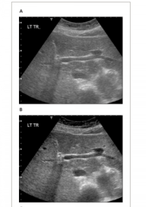
Here is an image that shows harmonics off (A) and on (B) for this particular case. Provided by Providian.

Comments
Thanks Diane for the detailed
Thanks Diane for the detailed explanation!
you’re welcome! I learned a
you’re welcome! I learned a good bit here myself by researching. And there is a lot more out there for those that are ultrasound nerds and love to get into the nitty-gritty!
Great stuff Diane!
Great stuff Diane!