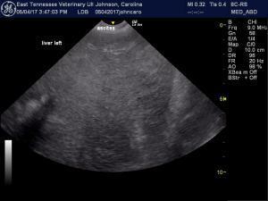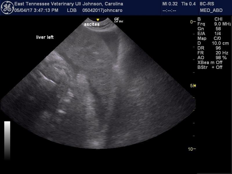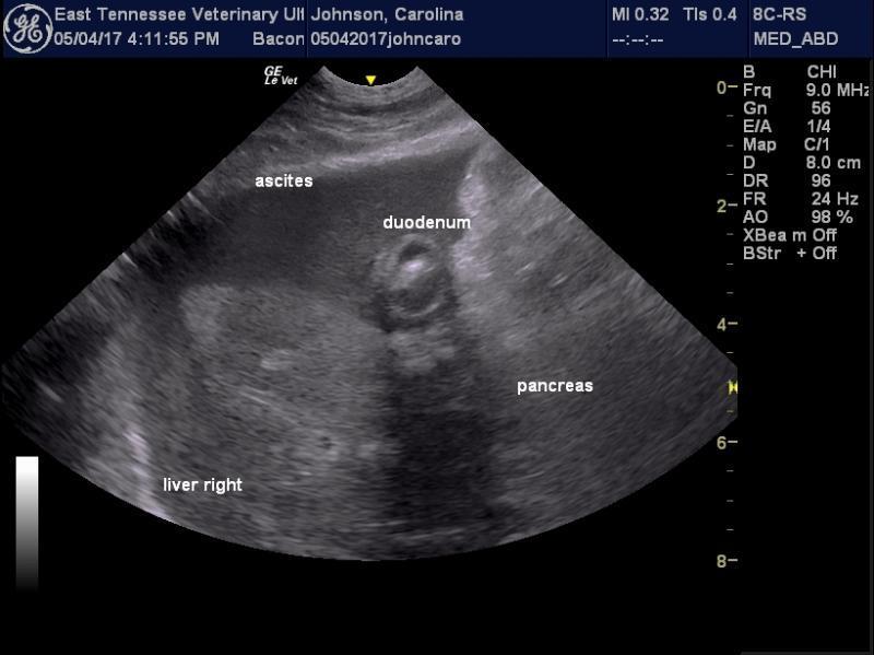

 These images were obtained from a 12 year old FS labrador who had been diagnosed with a gallbladder mucocele 2 years prior. Her owners declined surgery and she had been treated medically and did well until this past week when she developed acute vomiting, lethargy and weakness.
Her labwork showed moderate ALT and ALP elevations and mild bilirubinuia and bilirubinemia with WBC of 20 k. A CPL was normal.
Her images looked consistent with a gallbladder rupture and subsequent peritonitis. A centesis of fluid near the spleen revealed cloudy bright yellow fluid and what appeared (to me) to be numerous reactive mesethelial cells, neutrophils and some macrophages. No bile pigment was noted.
Further aspirates, including from regions near the gall bladder and liver, were declined and the patient was euthanized due to the severity of her clinical signs.
The clinician wasn’t convinced it was peritonitis due to the gallbladder rupture because there was not bile in the fluid we sampled. I observed that if it acute and/or walled off it may not show in the sample we obtained. The clinician was concerned with mesothelioma or carcinoma due to the cells we saw and I advised that reactive mesothelial cells can often have many criteria of malignancy and that a pathologist’s review would be beneficial.
Do you have any other thoughts? Another thought was regional peritonitis due to severe pancreatitis. I’ve never seen the inflammation appear like this at the gallbladder from that but suppose it is possible. Thank you!
Comments
I dont see the Gb here but
I dont see the Gb here but that amount of effusion and nodular hepatic changes I would be worried about neoplasia… i would fna liver and spleen and cytospin the fluid and analyze the sediment for neoplastic cells.
Hi Dr. Lindquist,
Did you see
Hi Dr. Lindquist,
Did you see the video? Thanks.
Thank you. Did you see the
Thank you. Did you see the video of the gallbladder?
yes I did but I cant make out
yes I did but I cant make out what is going on its a bit dark and the depth is minimal… would have to lower frequency, increase gain and depth to level of diaphragm and cover more wider sweep on the video as well as interrogate the portal hilus sweeping from cuystic duct region to gb… C-loop position 14 on the sdep protocol and position 12 right intercostal would be best here.