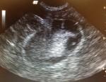
- A 12-year-old MN Cocker Spaniel mix with a host of issues presented for PTS.
- The dog has persistent liver enzyme elevations and all other blood work was significantly ABNORMAL to put it mildly.
- He also had a great deal of problems with his heart, was beginning to have cluster seizures; the list goes on.
- The owners opted for humane euthanasia, thankfully.? Sad as it was, it was definitely time.
- After the euthanasia I took the opportunity and asked the doctor if I could take a peek with the ultrasound probe, she said absolutely and watched me scan.
- I was actually just trying to practice finding my k9 organs, bladder/urethra, then kidney, (still can’t find my adrenal glands), spleen, and then…hello!
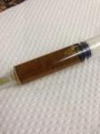
Image 3 is gallbladder sludge obtained via U/S-guided FNA (Are there any benefits in sampling the fluid in the gallbladder?)
Again my interest in putting a needle in it was purely for practice reasons, so I wasn’t sure if an FNA of the GB fluid would help diagnose anything.? I think the norm is to make sure the mucocele is not causing a problem, perf or other, but is not normally sampled.
Is this a gallbladder mucocele? It appeared pretty big and rather sludgy.? I compared my images with images from the pathology keyword/clinical search engine and they seem mighty similar.? http://www.sonopath.com/backroom/Secure/Search/StudyDetails.aspx?SID=406
Thanks in advance from all the pros out there!
Never pass up the opportunity to practice your scanning!? 🙂


Comments
Hey Sonogirl –
I have sampled the gallbladder before when I need to culture it, if I am concerned about chronic resistant infection. The recommendation is to empty it all the way to prevent leakage and subsequent bile peritonitis. I personally wouldn’t stick a mucocoele because you couldn’t empty it and the wall is often very friable anyway. I would worry about worsening an already bad situation. Since the animal is going to need surgery anyway, culture and sensitivity can be done at that time.
No expert, but that’s my opinion…
Liz
Hi Liz, thanks for the fast response. I normally would only stick bladders, but post-mortem was feeling brave. Is this a true GB mucocele? Or just a sludgy GB?
This is a mucocele, not a really bad or inflamed one but it is one. Overdistention, immobile suspended debris. I only stick gb if its more of a cholecystitis presentation with swirling mobile debris. GB mucoceles are like the worst toothpaste anal gland you have ever expressed so you don’t want that leaking in the cranial abdomen especially since the Gb wall in mucoceles is often fragile and likely to leak. look at our article and study on mucoceles Defining A Gall Bladder Mucocele. Number 2 for ecvim 2009 2. Clinical Parameters in Dogs with Sonographically Diagnosed Surgical Biliary Disease.
Thank you Dr. Lindquist, and sad to say I know the toothpaste you are speaking of, haha. The fluid I pulled off was very watery-not thickened at all but had tons of deep greenish flecks in it.
Likely more of an emerging mucocele then. Cockers are one of the top 3 breeds along with shelties and the terrier breeds to get mucoceles. German shepherd get them too but they are long football shaped. The mucocele seems to take the shape of the dog conformation:) Its just a challenge sometimes to tell if its clinically significant or not but they love, at the level of your mucocele here, to cause low grade signs. I have had cases where I just said “Looks like a mucocele but not inflamed but it isnt functional so lets get it out.” Then the owners say they have a new dog, vibrant and energetic. Don’t want to remove every sludgy Gb though but can confirm poor function wiht a GB motility study:
NPO, measure the Gb on US from right intercostal and subxyphoid approaches. Feed a/d or something fatty to stimulate CCK and Gb function and measure the Gb again at 15 and 30 min. If its not getting smaller its likely useless. Sharon center did unpublished work on this at Cornell.
Interesting, Eric – Simple study to do. I have had the same experience with “emerging mucocoeles,” where histo did not confirm mucocoele but dog feels 100% better after removal.
Yeh After being in the middle of this concept since the beginning I have come to the conclusion that “Mucocele” is a concept as opposed to a histopathological dx. Get it out & they feel better and its as systemically draining as a bad anal gland is chronically fastidious:)