 Lulu is a 6 year old F/S DSH feline that was presented to us yesterday with a 1 month period of wt loss. In that period of time she approximately lost half of her weight. In the last two days time she had suddenly become very lethargic and inappetent. On presentation she was not uremic and her coat was excellent. On manual abdominal palpation a firm and tortuous linear density was discovered. Since all of the changes seem to start after the previous holiday season a FB was a possibility. An abdominal ultrasound was performed to look for a cause of the wt loss.
The findings included the following;
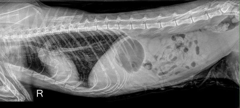
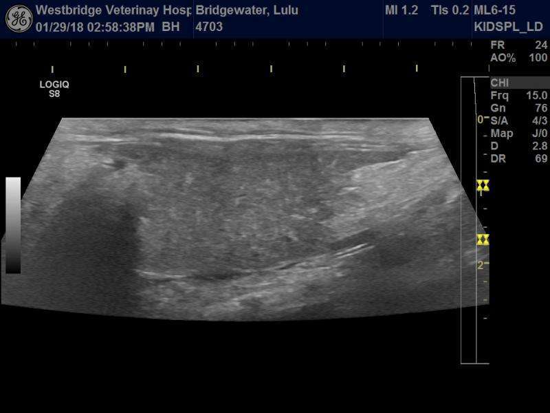
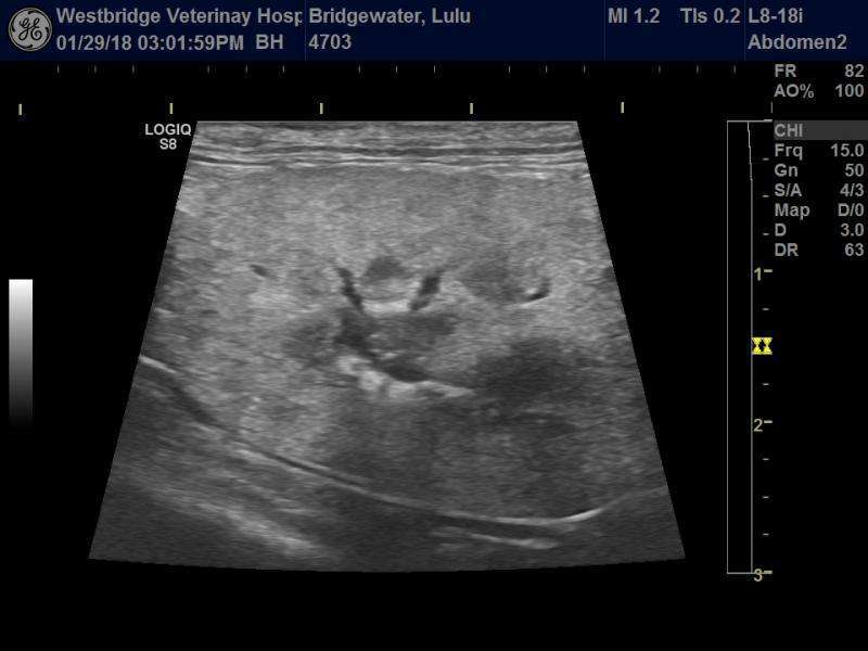
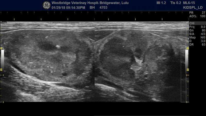
1) mineralization of the kidneys. This becomes even more obvious when some post mortem images where taken which accentuates the mineralization of the cortex.
2) mineralization within the spleen
3) enlarged splenic and portal veins
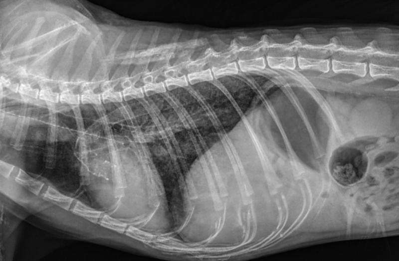
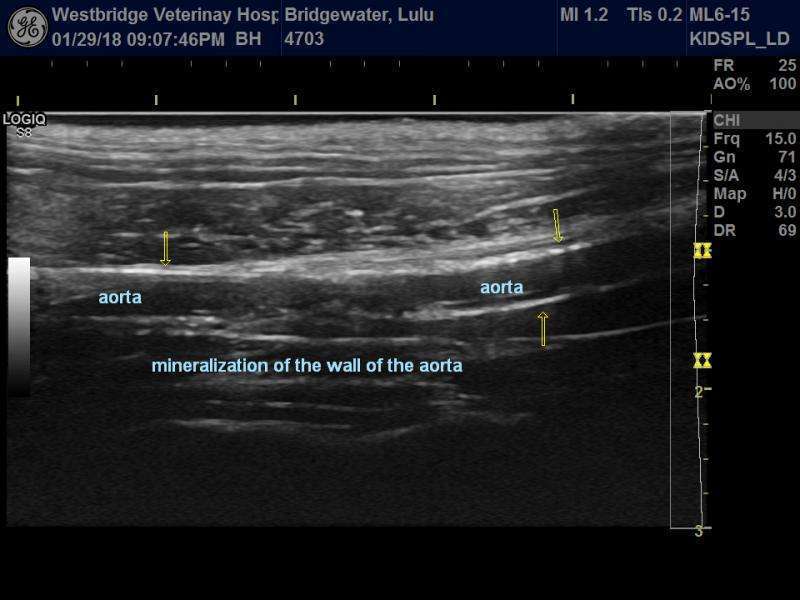
4) mineralized dorsal aorta and aortic trunk
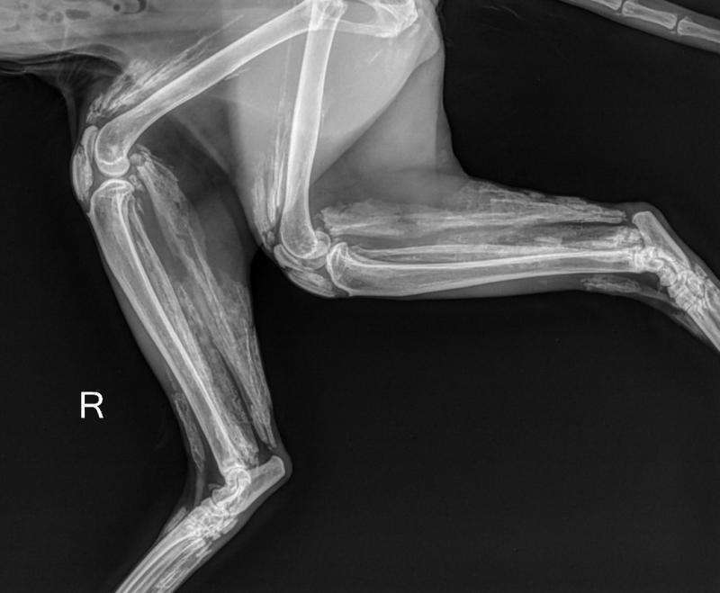
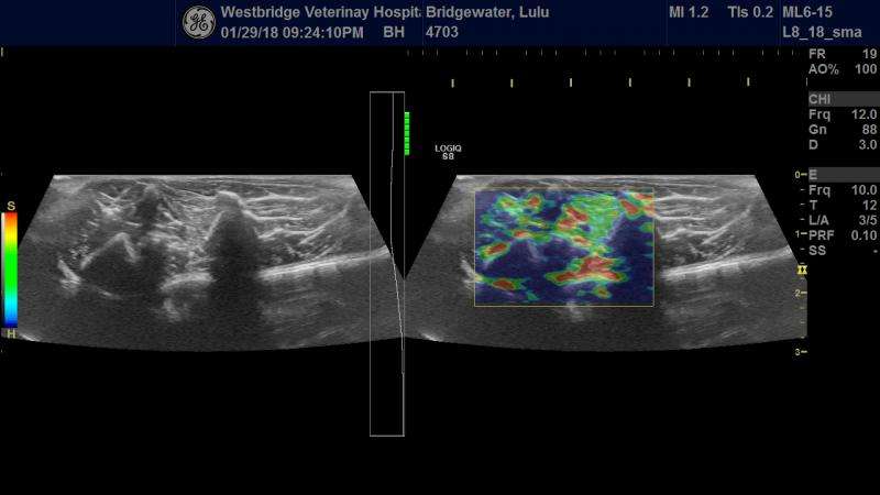
5) the distal tendinous portions of many muscles are mineralized as well as much of the cardiac myocardium
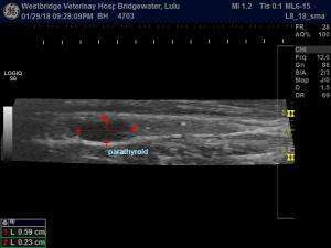
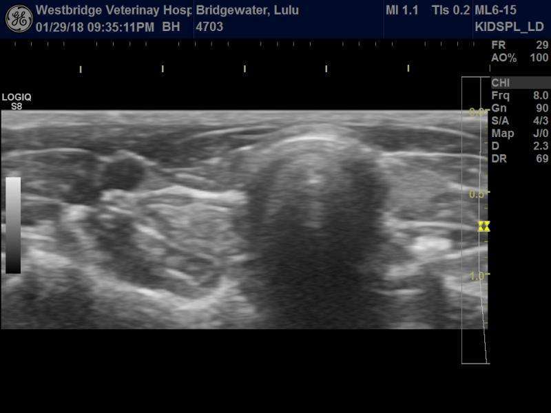
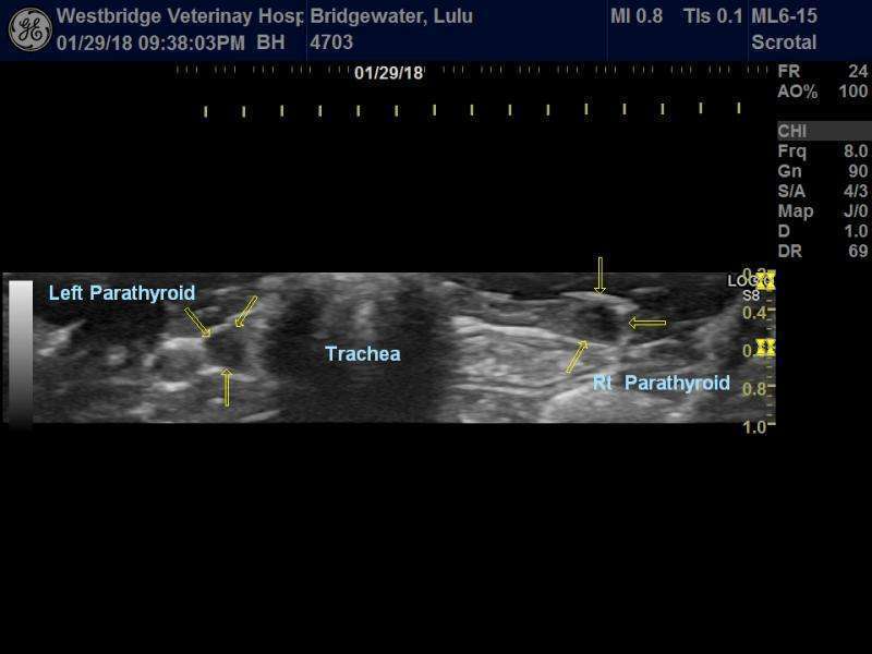
6) Both parathyroid glands are very prominent and measure 2.6 X 2.2 X 5.9mm
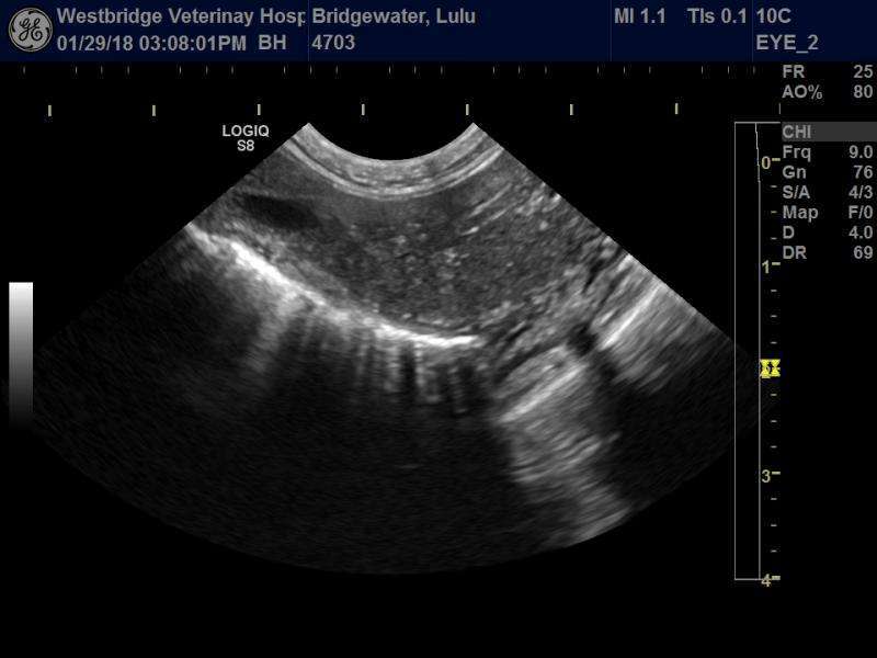
7) mineralization of the bronchial tree. Many rockets on the pulmonary ultrasound.
These are all dramatic changes that are both seen on Ultrasound and radiology. The owner was not aware of any mobility issues prior to a few days ago. Unfortunately we do not have serum renal biochemistry values or any ionized calcium values. Neither do we have PTH levels. The owner rightfully opted for E/D.
The question is whether this is primary parathyroid disease or more as is supected secondary to chronic renal disease. As I mentioned I suspect that this is secondary to the renal disease but two things bother me considering that we are missing blood values;
a) if it was renal in nature then why was there no azotemia on physical examination when smelling her breath?
b) I know that parathyroid masses should be unilateral but then why are they so prominent. They are way to easy to image and in light that I cannot find references to normal measurements. Maybe they are normal and I am just lucky illustrating them. Any thoughts?
Comments
Beautiful image set as always
Beautiful image set as always Bob. Since you are new to the forum I want to just let you know that forum protocol is a few stills and a few videos per thread so we can all move through them quickly in our busy days otherwise it becomes a telemed platform which isnt its purpose. Please see the forum rule description in the upper left.
That being said this is a very compelx case and I can see the impetus to spend the time to upload so many so we cvan all nerd up appropriately so I thank you for that:)
This is truly a really cool case. Honestly I would FNA those thyroid nodules and send to a trusted cytologist… mine is in sonopath telecytology Larry McGill (plug plug) but when you have a cytologist you trust you build a service around him/her so I did. 25 or 27g fna to differentiate adenoma from hyperplasia and if adenoma then cut it out… that is in vivo ideally.
If singular nodule/gland even bilaterally I would bet on adenoma as renal hyperparathyroidism causes multiple nodules of various size in my experience but Nele is the small parts queen so Ill check with her but shes in a cluster of university, teleradiology and lectures/conference the next couple of days so likely will be a delay. But the less experience I have with something the faster I go to a needle to enhance my curve and knowledge and would have gone there on this case mainly becaus ei fdont liek to think this much and a good needle keeps me from getting a head scratching headache:)
Also I see functional non renal failure kidneys look like this all the time with interstital nephrosis pattern of course not with this pattern of cortical mineralization but more cm mineralization… cat kidneys can really get lumped up before smoking 65% of the functionality to enter into renal failure.
if you had parathyroid histopath its of publishable quality I think.
Eric, my apologies. I did not
Eric, my apologies. I did not mean to tie up your time. I just thought tht it was an unusual case that could offer a teaching moment. I will respect the rules in the future. I honestly wasn’t aware of the listed submission rules in red. Thank you for the heads up.
The question still remains though; does anyone have a range for the normal size of parathyroids in felines?. If these are way off base then it almost answers the question of who done it. Overly big glands …….probably adenomas. Normal size….. then more probably renal in origin.
I could not biopsy the glands. Owner decided to euthanize the poor cat since we would never be able to significantly reverse most of the soft tissue changes. She was very pregnant and very very emotional. I wasn’t going to ask her for permission to aspirate the nodes post euthanasia. I honestly didn’t want to precipitate a miscarriage.
I don’t mind being a nerd:). As for publishing I am working on the adrenal US and Cushing’s disease study first. I hope to have it done by the summer. We will see what the future brings.
>>I fdont like to think this much and a good needle keeps me from getting a head scratching headache:)<<<
Why do you think that I’m bald!
No worries Bob I just have to
No worries Bob I just have to be the forum cop on occasion:) We could stay on the forum all day long but wouldnt get anything done in our normal lives so I designed the forum to boom boom, pop in, add a piece to the thread and everyone keeps going… bullet point style. I learned this from other forums as you know can be exhaustive to sift through so we want to keep it tight, stimulate thoughts, add a thought or 2, and move on with our work flows.
If anyone has the normals on feline parathyroids Nele would. Lets see what she says after her seminar this weekend. Looking forward to your adrenal study there isnt enough info on that subject in publication for sure.
With this extensive
With this extensive minerlisation/calcification is there any possiblity of exposure to the calcium containing rodenticides or owners calcium supplements/calcitriol?
None what so ever on both
None what so ever on both counts
Not sure how applicable this
Not sure how applicable this is:
An atypical case of severe soft–tissue mineralization in a 3-week-old foal from a herd of Andalusian horses. The herd clinical history and the laboratory findings were compatible with a diagnosis of secondary hyperparathyroidism due to a mineral imbalance in the diet (low calcium and high phosphorus intake). Mares showed a marked increase in serum parathyroid hormone (PTH) approximately 10 times normal levels. Serum PTH was marginally elevated in foals. Clinical signs (unthriftiness, painful joints, lameness in one or more limbs, and stiff gait) were more pronounced in foals than in mares. Two foals died and necropsy of one of them revealed extensive soft–tissue mineralization of arterial walls and pulmonary parenchyma. Clinical signs in mares and foals resolved by 4 weeks after diet adjustment.
And getting to the bottom of
And getting to the bottom of the barrel :))
A 26-year-old woman with systemic lupus erythematosus and extensive soft tissue calcification.
Those cases are truly
Those cases are truly fascinating. I will inquire about the diet the owner was feeding. You just never know until you ask. Something is amiss here.. I could not get very far along with this case as I would have wished for. The owner pretty well made the decision to stop once she saw the rads. This one will pull at my curiosity for some time. I hope to have the parathyroid normal measurements very soon.
In an ideal world we would
In an ideal world we would have full bloods and hormone assays, which would made it a bit easier to diagnose :))
True but I’m still kicking
True but I’m still kicking myself.
It appears that the normal
It appears that the normal measurement as per Mark Peterson is a 4 mm round but flat stucture. The second parathyroid is embeded in the thyroid gland and cannot be seen on US.
If on a longitudinal ( sagittal) view it should be visualized as being round then the diameter should have been 2.3 mm and not 5.9mm as per these images. The thickness should have been less then 2.3 mm to be flat if we follow the same logic. They are not flat on these images unless the “flatness” is expected to be on the sagittal view. Either way they are too large. So I am to believe that these are secreting adenomas.
Nele will have the final word on whether these are normal or not.