Hello
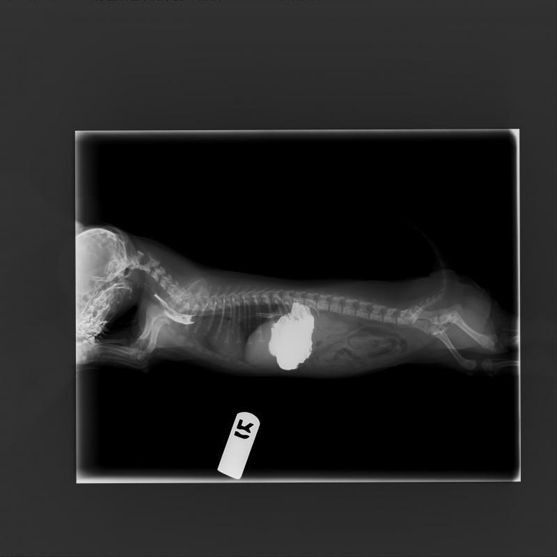
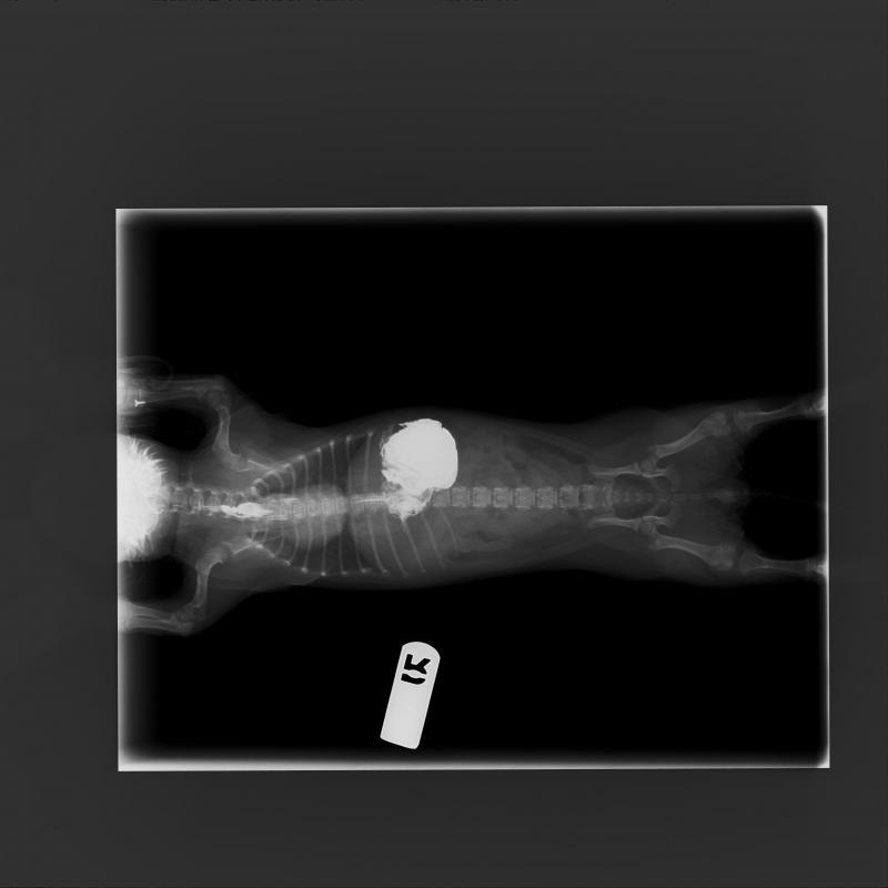
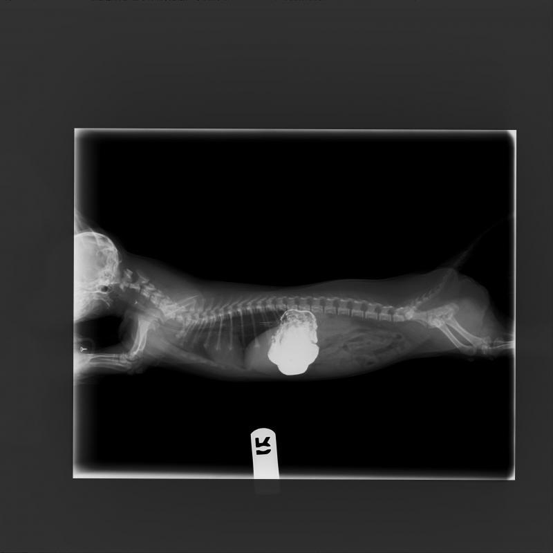
This is Angel an 8 week old Shi Tzu cross that presented to us for dysphagia, salivation. The referring veterinarian took scout rads and thought there might be an esophageal fb and sent it to us to scope. However because of the size (5lbs) we were not able to so elected for barium swallow instead.
At T1 there is a mineral opacity overlying the trachea seen on regular DVM rads and can be seen on ours after barium has passed. This should be the 4th image and the teaser image.
There appears to be a filling defect in that area but does not appear to be related to mineral opacity as it is lateral to the esophgus and does not retain any barium.
I was hoping for some opinions on what the filling defect could be. Esophageal FB vs congenital lesion vs open. Angel has started eating and drinking well and is clinically improved so we are taking a conservative approach. We have mentioned CT but I would like some opinions before we consider proceeding on a clinically well dog.
Thanks. Brent.


Comments
I think its too cranial for a
I think its too cranial for a PRAA …OPther than let him grow into the lesion and then scope or let it be more dramatic on rads. I’m deferring to Nele on this one:)
That was out plan!
To scope
That was out plan!
To scope in a few months if still present. Thanks For the comments!